 Indietro
Muscles and Muscle Tissue: Structure, Function, and Physiology
Guida di studio - Note intelligenti
 Appunti personalizzati basati sui tuoi materiali, ampliati con definizioni chiave, esempi e contesto.
Muscle Tissue Overview
Introduction to Muscle Tissue
Muscle tissue is essential for movement, posture, and various bodily functions. It comprises nearly half of the body's mass and is specialized to convert chemical energy (ATP) into mechanical energy, producing force and movement.
Key Terminology: Prefixes such as myo-, mys-, and sarco- refer to muscle (e.g., sarcoplasm is muscle cell cytoplasm).
Three Types of Muscle Tissue: Skeletal, Cardiac, and Smooth.
Types of Muscle Tissue
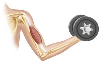
Skeletal Muscle: Attached to bones and skin, responsible for voluntary movements, striated, and multinucleate. Contracts rapidly but tires easily.
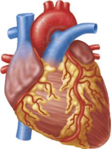
Cardiac Muscle: Found only in the heart, striated, involuntary, and contracts at a steady rate due to the heart's pacemaker.
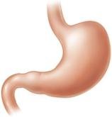
Smooth Muscle: Located in walls of hollow organs (e.g., stomach, bladder, airways), non-striated, involuntary, and spindle-shaped.
Comparison of Muscle Types
Characteristic | Skeletal | Cardiac | Smooth |
|---|---|---|---|
Location | Bones/skin | Heart walls | Walls of hollow organs |
Cell Shape | Long, cylindrical, multinucleate, striated | Branching chains, uni/binucleate, striated | Spindle-shaped, uninucleate, non-striated |
Control | Voluntary | Involuntary | Involuntary |
Characteristics of Muscle Tissue
Excitability: Ability to receive and respond to stimuli.
Contractility: Ability to shorten forcibly when stimulated.
Extensibility: Ability to be stretched.
Elasticity: Ability to recoil to resting length.
Functions of Muscle Tissue
Produce movement (locomotion, manipulation, pumping blood, digestion)
Maintain posture and body position
Stabilize joints
Generate heat during contraction
Skeletal Muscle Structure and Organization
Components of Skeletal Muscle
Skeletal muscle is an organ composed of muscle fibers, connective tissue, nerves, and blood vessels. Each muscle receives a nerve, artery, and veins, ensuring control and nutrient supply.
Connective Tissue Sheaths
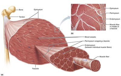
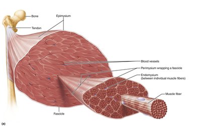
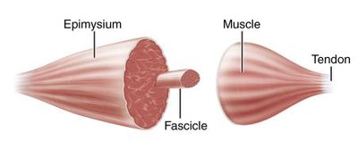
Epimysium: Dense irregular connective tissue surrounding the entire muscle; may blend with fascia.
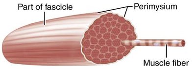
Perimysium: Fibrous connective tissue surrounding fascicles (bundles of muscle fibers).
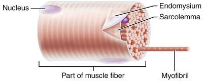
Endomysium: Fine areolar connective tissue surrounding each muscle fiber.
Muscle Attachments
Direct (fleshy) Attachments: Epimysium fused to periosteum of bone or perichondrium of cartilage.
Indirect Attachments: Connective tissue wrappings extend beyond muscle as a tendon or aponeurosis.
Structural Organization
Level | Description | Connective Tissue |
|---|---|---|
Muscle (organ) | Hundreds to thousands of muscle cells, blood vessels, nerves | Epimysium |
Fascicle | Bundle of muscle cells | Perimysium |
Muscle fiber (cell) | Elongated, multinucleate, striated cell | Endomysium |
Microscopic Anatomy of Skeletal Muscle Fibers
Muscle Fiber Structure
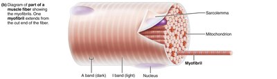
Sarcolemma: Plasma membrane of muscle fiber.
Sarcoplasm: Cytoplasm containing glycosomes (glycogen storage) and myoglobin (O2 storage).
Specialized Structures: Myofibrils, sarcoplasmic reticulum, and T tubules.
Myofibrils and Sarcomeres
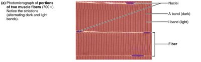
Myofibrils: Densely packed, rodlike elements making up ~80% of cell volume. Responsible for striations.
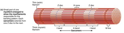
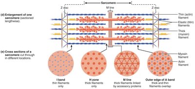
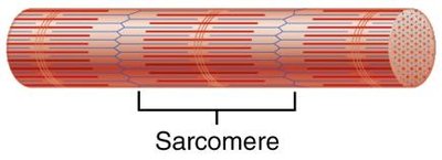
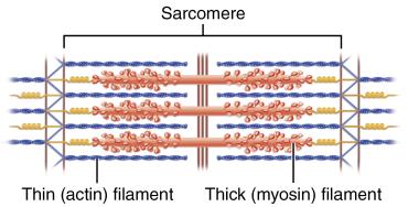
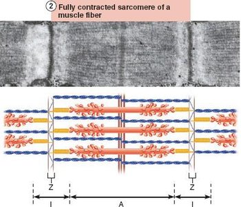
Sarcomere: Smallest contractile unit, defined as the region between two Z discs. Contains A band (dark) and I band (light).
Myofilaments
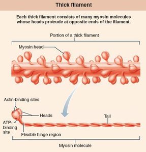
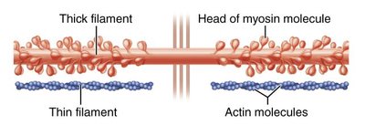
Thick Filaments: Composed of myosin molecules with heads that form cross bridges during contraction.
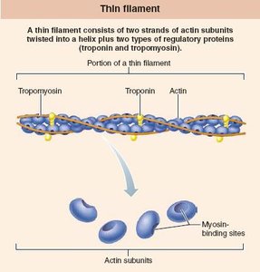
Thin Filaments: Composed of actin, tropomyosin, and troponin. Actin subunits provide binding sites for myosin heads.
Elastic Filament: Composed of titin, helps maintain sarcomere structure and elasticity.
Dystrophin: Links thin filaments to sarcolemma proteins.
Clinical Application: Duchenne Muscular Dystrophy (DMD)
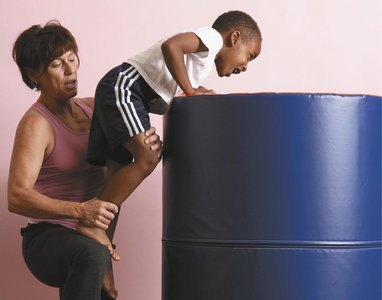
DMD is a severe, inherited muscle-destroying disease caused by a defective gene for dystrophin. It leads to progressive muscle weakness and loss of muscle mass, primarily affecting males. There is no cure, but supportive care and gene therapy can extend life expectancy.
Sarcoplasmic Reticulum and T Tubules
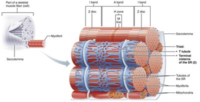
Sarcoplasmic Reticulum (SR): Smooth ER network surrounding each myofibril, stores and releases Ca2+ for contraction.
T Tubules: Invaginations of the sarcolemma that conduct electrical impulses deep into the muscle fiber, ensuring coordinated contraction.
Triad: Structure formed by a T tubule and two terminal cisterns of the SR.
Sliding Filament Model of Contraction
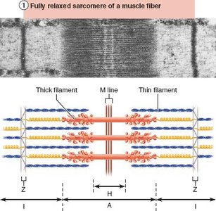
The sliding filament model explains how muscles contract: thin filaments slide past thick filaments, increasing their overlap without changing filament length. Cross bridges form and break, pulling thin filaments toward the center of the sarcomere, resulting in muscle shortening.
Neuromuscular Junction and Muscle Contraction
Excitation of Muscle Fibers
Motor neurons transmit signals from the brain/spinal cord to muscle fibers.
Action potentials (APs) travel along neurons, leading to the release of acetylcholine (ACh) at the neuromuscular junction (NMJ).
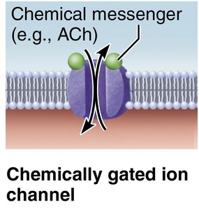
ACh binds to receptors on the sarcolemma, opening chemically gated ion channels and initiating muscle fiber excitation.
Steps in Muscle Fiber Excitation
AP arrives at axon terminal.
Voltage-gated Ca2+ channels open; Ca2+ enters neuron.
Ca2+ triggers ACh release into synaptic cleft.
ACh binds to sarcolemma receptors, opening Na+ channels (end plate potential).
AP propagates along sarcolemma and T tubules.
ACh is degraded by acetylcholinesterase.
Excitation-Contraction Coupling and Cross Bridge Cycling
Excitation-Contraction (E-C) Coupling
AP travels along sarcolemma and down T tubules.
Voltage-sensitive proteins trigger Ca2+ release from SR.
Ca2+ binds to troponin, moving tropomyosin and exposing actin binding sites.
Myosin heads bind to actin, initiating contraction.
Cross Bridge Cycle
Cross bridge formation: Myosin head attaches to actin.
Power stroke: Myosin head pivots, pulling actin filament toward M line.
Cross bridge detachment: ATP binds to myosin, causing detachment.
Cocking of myosin head: ATP hydrolysis re-energizes myosin head.
Whole Muscle Contraction
Motor Units
A motor unit consists of a motor neuron and all the muscle fibers it innervates.
Smaller motor units allow for fine control; larger units generate more force.
Muscle Twitch and Graded Responses
Muscle Twitch: Response to a single AP; consists of latent, contraction, and relaxation phases.
Graded Muscle Contractions: Strength and duration of contraction can be varied by changing stimulus frequency (temporal summation) or strength (recruitment).
Isotonic vs. Isometric Contractions
Isotonic: Muscle changes length (shortens or lengthens) and moves a load.
Isometric: Muscle tension increases but does not exceed load; muscle does not change length.
ATP and Muscle Metabolism
ATP Sources for Contraction
Direct Phosphorylation: Creatine phosphate donates phosphate to ADP to form ATP.
Anaerobic Pathway: Glycolysis and lactic acid formation (no oxygen required).
Aerobic Pathway: Glycolysis followed by aerobic respiration in mitochondria (requires oxygen).
Equation for Direct Phosphorylation:
Muscle Fatigue and Recovery
Muscle Fatigue: Inability to contract despite stimulation, often due to ionic imbalances, increased inorganic phosphate, or decreased ATP.
Excess Postexercise Oxygen Consumption (EPOC): Extra oxygen needed to restore muscle to resting state after exercise.
Factors Affecting Muscle Contraction
Frequency of stimulation
Number of muscle fibers recruited
Size of muscle fibers
Degree of muscle stretch (length-tension relationship)
Muscle Fiber Types
Type | Speed | ATP Pathway | Fatigue Resistance | Best Suited For |
|---|---|---|---|---|
Slow Oxidative | Slow | Aerobic | High | Endurance, posture |
Fast Oxidative | Fast | Aerobic (some anaerobic) | Intermediate | Sprinting, walking |
Fast Glycolytic | Fast | Anaerobic glycolysis | Low | Short, intense movements |
Smooth Muscle
Structure and Function
Found in walls of hollow organs (except heart).
Spindle-shaped, uninucleate, non-striated cells.
Organized in longitudinal and circular layers for coordinated contractions.
Differences from Skeletal Muscle
Only endomysium connective tissue present.
Innervated by autonomic nervous system via varicosities (diffuse junctions).
SR less developed; Ca2+ mainly from extracellular fluid via caveolae.
Gap junctions allow electrical coupling between cells.
Thick filaments have myosin heads along entire length; no troponin (calmodulin binds Ca2+).
Intermediate filaments and dense bodies anchor actin filaments.
Contraction Mechanism
Slow, synchronized contractions; some cells act as pacemakers.
Ca2+ binds to calmodulin, activating myosin kinase, which phosphorylates myosin for cross bridge formation.
Relaxation requires Ca2+ detachment, active transport, and myosin dephosphorylation.
Types of Smooth Muscle
Unitary (Visceral): Found in hollow organs, electrically coupled, spontaneous APs, responds to chemical stimuli.
Multi Unit: Found in large airways, arteries, arrector pili, iris; independent fibers, graded contractions, autonomic control.
Development and Aging of Muscle Tissue
All muscle tissues develop from embryonic mesoderm cells called myoblasts.
Skeletal muscle cells form by fusion of myoblasts; cardiac and smooth muscle cells do not fuse but develop gap junctions.
Muscle regeneration is limited; smooth muscle regenerates throughout life, but skeletal and cardiac muscle have limited capacity.
With aging, muscle mass decreases (sarcopenia), but regular exercise can slow this process.
