 Indietro
Muscles and Muscle Tissue: Structure, Function, and Organization
Guida di studio - Note intelligenti
 Appunti personalizzati basati sui tuoi materiali, ampliati con definizioni chiave, esempi e contesto.
Muscle Tissue Characteristics
Major Functions of Muscle Tissue
Muscle tissue is essential for movement, posture, joint stabilization, and heat generation. Nearly half of the body’s mass is muscle, which transforms chemical energy (ATP) into mechanical energy to exert force.
Produce movement: Responsible for locomotion and manipulation (e.g., walking, digesting, pumping blood).
Maintain posture and body position: Muscles continuously adjust to keep the body upright.
Stabilize joints: Muscles reinforce and stabilize joints during movement.
Generate heat: Muscle contraction produces heat, helping maintain body temperature.
Characteristics Shared by All Muscle Tissue
Excitability (Responsiveness): Ability to receive and respond to stimuli.
Contractility: Ability to shorten forcibly when stimulated.
Elasticity: Ability to recoil to resting length after stretching.
Extensibility: Ability to be stretched beyond resting length.
Types of Muscle Tissue
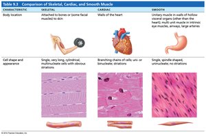
Comparison of Skeletal, Cardiac, and Smooth Muscle
There are three types of muscle tissue in the human body, each with distinct anatomy, function, and distribution.
Skeletal Muscle: Attached to bones or skin; voluntary control; striated; single, long, cylindrical, multinucleate cells.
Cardiac Muscle: Found in the heart; involuntary control; striated; branching chains of cells; usually one nucleus per cell.
Smooth Muscle: Found in walls of hollow organs (except heart); involuntary control; non-striated; single, spindle-shaped cells with one nucleus.
Characteristic | Skeletal | Cardiac | Smooth |
|---|---|---|---|
Body Location | Attached to bones or skin | Walls of the heart | Walls of hollow organs |
Cell Shape & Appearance | Long, cylindrical, striated, multinucleate | Branching, striated, one or two nuclei | Spindle-shaped, non-striated, one nucleus |
Skeletal Muscle Anatomy
Gross Structure of Skeletal Muscle
Skeletal muscle is an organ composed of muscle fibers, connective tissue, nerves, and blood vessels. It is highly vascularized and innervated, ensuring efficient delivery of nutrients and removal of waste.
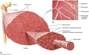
Muscle fascicle: Bundle of muscle fibers surrounded by connective tissue.
Skeletal muscle fiber (myocyte): Elongated, multinucleate cell; basic unit of muscle contraction.
Myofibril: Rod-like structure within muscle fiber, composed of repeating units called sarcomeres.
Myofilament: Protein filaments (actin and myosin) within myofibrils responsible for contraction.
Connective Tissue Layers Surrounding Skeletal Muscle
Epimysium: Surrounds entire muscle.
Perimysium: Surrounds each fascicle.
Endomysium: Surrounds individual muscle fibers.
Muscle Fiber Anatomy
Organization and Structures within a Skeletal Muscle Fiber
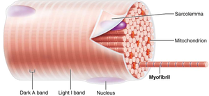
Each muscle fiber contains specialized structures for contraction and regulation.
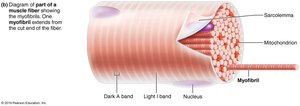
Sarcolemma: Plasma membrane of the muscle cell.
Sarcoplasm: Cytoplasm of the muscle cell, containing organelles and myofibrils.
Sarcoplasmic reticulum (SR): Specialized endoplasmic reticulum that stores and releases calcium ions (Ca2+).
Transverse tubules (T-tubules): Invaginations of the sarcolemma that transmit electrical signals deep into the muscle fiber.
Myofibrils: Cylindrical structures composed of sarcomeres.
Myofilaments: Actin (thin), myosin (thick), titin (elastic), troponin, and tropomyosin.
Sarcomere Structure and Myofilament Composition
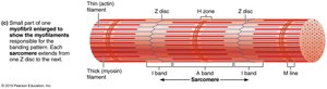
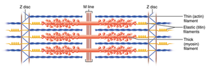
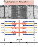
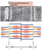
The sarcomere is the functional unit of striated muscle, defined by the region between two Z-discs. It contains overlapping thick and thin filaments, which create the banding pattern observed in muscle fibers.
A-band: Dark region containing thick (myosin) filaments.
I-band: Light region containing thin (actin) filaments.
H-zone: Central region of A-band with only thick filaments.
Z-disc (Z-line): Boundary of the sarcomere; anchors thin filaments.
M-line: Center of the sarcomere; anchors thick filaments.
Proteins in the Sarcomere
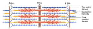
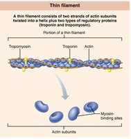
Actin: Forms thin filaments; contains myosin-binding sites.
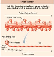
Myosin: Forms thick filaments; has heads that bind to actin and ATP.
Titin: Elastic protein that stabilizes thick filaments and allows recoil.
Troponin and Tropomyosin: Regulatory proteins on thin filaments.
Dystrophin: Links thin filaments to sarcolemma proteins.
Nebulin, Myomesin, C proteins: Maintain sarcomere alignment.
Sarcoplasmic Reticulum and T Tubules
Relationship to Myofibrils
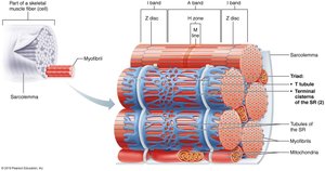
The sarcoplasmic reticulum (SR) and T tubules are critical for excitation-contraction coupling. The SR stores calcium ions, which are released upon stimulation. T tubules transmit electrical impulses from the sarcolemma to the SR, triggering calcium release.
Terminal cisterns: Enlarged regions of SR at the A–I band junction.
Triad: Structure formed by a T tubule and two terminal cisterns.
Voltage sensors: Proteins in T tubules that respond to electrical signals.
Calcium channels: Proteins in SR that release Ca2+ when activated.
Changes in Sarcomere Size During Muscle Contraction
Sliding Filament Model of Contraction
Muscle contraction occurs when myosin heads bind to actin, pulling the thin filaments toward the center of the sarcomere. This shortens the sarcomere, resulting in muscle contraction.
Relaxed sarcomere: A-band remains constant; I-band and H-zone are wider.
Contracted sarcomere: A-band remains constant; I-band and H-zone narrow or disappear.
Summary Table: Structure and Organizational Levels of Skeletal Muscle
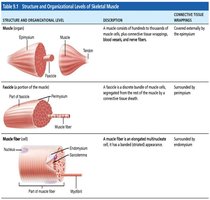
Structure and Organizational Level | Description | Connective Tissue Wrapping |
|---|---|---|
Muscle (organ) | Hundreds to thousands of muscle cells, plus connective tissue, blood vessels, and nerve fibers | Epimysium |
Fascicle (portion of muscle) | Discrete bundle of muscle cells, segregated from rest of muscle by connective tissue sheath | Perimysium |
Muscle fiber (cell) | Elongated multinucleate cell; has striated appearance | Endomysium |
Key Terms and Definitions
Sarcolemma: Muscle cell plasma membrane.
Sarcoplasm: Muscle cell cytoplasm.
Sarcoplasmic reticulum: Specialized ER for Ca2+ storage and release.
Myofibril: Rod-like contractile element within muscle fiber.
Myofilament: Actin (thin) and myosin (thick) filaments.
Sarcomere: Functional unit of muscle contraction.
Equations and Formulas
ATP Hydrolysis: Muscle contraction is powered by ATP hydrolysis.
Sliding Filament Model: Sarcomere shortening is proportional to muscle contraction.
Example: Application of Muscle Tissue Types
Skeletal muscle: Used for voluntary movements such as walking and lifting.
Cardiac muscle: Responsible for pumping blood throughout the body.
Smooth muscle: Controls involuntary movements such as digestion and blood vessel constriction.
Additional info: Academic context was added to clarify the structure and function of muscle tissue, sarcomere organization, and the sliding filament model. Tables were recreated and expanded for clarity.
