 Indietro
Muscles and Muscle Tissue: Structure, Function, and Physiology
Guida di studio - Note intelligenti
 Appunti personalizzati basati sui tuoi materiali, ampliati con definizioni chiave, esempi e contesto.
Muscle Tissue Overview
Introduction to Muscle Tissue
Muscle tissue constitutes nearly half of the human body's mass and is essential for movement, posture, and heat generation. Muscles transform chemical energy (ATP) into mechanical energy, enabling forceful contractions and movement.
Types of muscle tissue: Skeletal, Cardiac, Smooth
Characteristics: Excitability, Conductivity, Contractility, Elasticity, Extensibility
Functions: Movement, posture, joint stabilization, heat production

Types of Muscle Tissue
Classification and Terminology
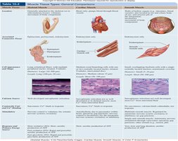
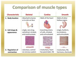
Muscle tissue is classified based on structure, function, and location. Prefixes such as myo, mys, and sarco are commonly used in muscle terminology (e.g., sarcoplasm is muscle cell cytoplasm).
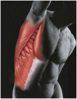
Skeletal muscle: Voluntary, striated, attached to bones and skin
Cardiac muscle: Involuntary, striated, found only in the heart
Smooth muscle: Involuntary, non-striated, found in walls of hollow organs
Characteristics of Muscle Tissue
Functional Properties
Muscle tissue exhibits several key properties that enable its function:
Excitability: Ability to respond to stimuli by changing membrane potential
Conductivity: Transmission of electrical impulses along the cell membrane
Contractility: Ability to shorten forcibly when stimulated
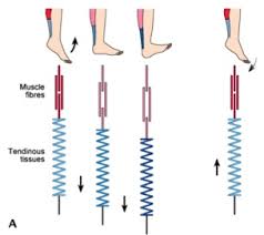
Elasticity: Ability to return to original length after stretching
Extensibility: Ability to be stretched beyond resting length
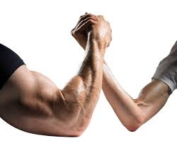
Muscle Functions
Major Roles of Muscle Tissue
Muscles perform four primary functions in the body:
Produce movement: Locomotion and manipulation (e.g., walking, digesting, pumping blood)
Maintain posture: Stabilize body position
Stabilize joints: Support and reinforce joints
Generate heat: Contraction produces heat, aiding in temperature regulation
Nerve and Blood Supply
Innervation and Vascularization
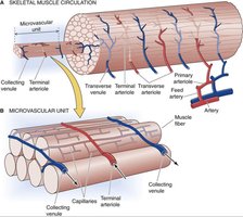
Each muscle receives a nerve, artery, and veins, which enter centrally. Skeletal muscle fibers require substantial oxygen and nutrients, and waste removal is essential for function.
Every muscle fiber is innervated for precise control
Blood supply supports metabolic needs
Muscle Attachments
Origin, Insertion, and Types of Attachments
Muscles span joints and attach to bones at two main points:
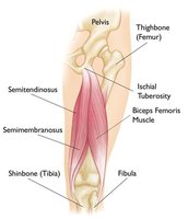
Origin: Attachment to immovable or less movable bone
Insertion: Attachment to movable bone
Direct attachment: Epimysium fused to periosteum or perichondrium
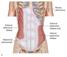
Indirect attachment: Connective tissue extends as tendon or aponeurosis (most common)
Connective Tissue Sheaths
Muscle Structure and Support
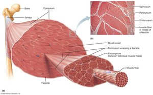
Connective tissue sheaths reinforce and support muscle structure:
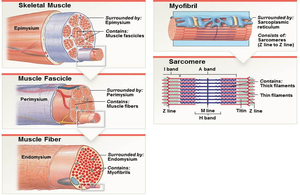
Epimysium: Surrounds entire muscle
Perimysium: Surrounds fascicles (bundles of muscle fibers)
Endomysium: Surrounds individual muscle fibers
All three contribute to tendon formation.
Structural Organization of Skeletal Muscle
Hierarchy of Muscle Structure
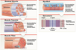
Skeletal muscle is organized into five hierarchical levels:
Whole muscle: Contains many fascicles
Fascicle: Bundle of muscle fibers
Muscle fiber (cell): Long, multinucleated cell
Myofibril: Rodlike unit within muscle fiber
Myofilaments: Actin (thin) and myosin (thick) filaments
Muscle Fiber Anatomy
Components of Muscle Cells
Skeletal muscle fibers are specialized cells containing:
Sarcolemma: Plasma membrane
Sarcoplasm: Cytoplasm
Myofibrils: Contain sarcomeres
Sarcoplasmic reticulum: Stores calcium
T tubules: Conduct action potentials
Myofibril and Sarcomere Structure
Organization and Function
Myofibrils are densely packed structures within muscle fibers, containing thousands of sarcomeres—the functional units of contraction.
Striations: Alternating dark (A band) and light (I band) regions
Sarcomere: Area between Z discs, smallest contractile unit
Myofilaments: Actin (thin) and myosin (thick) filaments
Molecular Composition of Myofilaments
Proteins and Their Functions
Myofilaments are composed of several proteins:
Myosin: Thick filament, forms cross bridges during contraction
Actin: Thin filament, contains binding sites for myosin
Tropomyosin: Covers myosin-binding sites on actin
Troponin: Regulatory protein, binds calcium
Titin: Elastic filament, maintains structure
Dystrophin: Links thin filaments to sarcolemma
Sliding Filament Model of Contraction
Mechanism of Muscle Contraction
Muscle contraction occurs when thin filaments slide past thick filaments, increasing overlap and shortening the sarcomere. Cross bridge formation and cycling are essential for this process.
Shortening: Z discs move closer, I bands shorten, H zones disappear
Cross bridge cycle: Myosin heads attach, pivot, detach, and reset
Clinical Relevance: Duchenne Muscular Dystrophy
Pathophysiology and Symptoms
Duchenne muscular dystrophy (DMD) is a sex-linked recessive disorder caused by defective dystrophin. It leads to muscle degeneration, weakness, and loss of function, primarily affecting males.
Symptoms: Clumsiness, frequent falls, progressive muscle weakness
Pathology: Sarcolemma tears, calcium influx, inflammation, apoptosis
Muscle Contraction: Four Steps
Sequence of Events
Four steps are required for skeletal muscle contraction:
Events at the neuromuscular junction: Release of acetylcholine (ACh)
Events at the sarcolemma: Muscle fiber excitation
Events at the T-tubule: Excitation-contraction coupling, Ca2+ release
Crossbridge cycling: Myosin-actin interaction
Motor Unit
Definition and Function
A motor unit consists of a motor neuron and all the muscle fibers it innervates. The number of fibers per motor unit determines the precision of control.
Fine control: Few fibers per motor unit
Strength: Many fibers per motor unit
Types of Muscle Contraction
Isometric vs. Isotonic
Muscle contractions are classified as:
Isometric: Muscle generates tension without changing length
Isotonic: Muscle changes length and moves load
Concentric: Muscle shortens
Eccentric: Muscle lengthens
Muscle Tone
Definition and Importance
Muscle tone is the constant, slightly contracted state of all muscles, maintained by spinal reflexes. It keeps muscles firm, healthy, and ready to respond.
Supplying Energy for Muscle Contraction
ATP Generation Pathways
Muscle cells generate ATP through three main pathways:
Immediate supply: Phosphate transfer (creatine kinase pathway)
Short-term supply: Glycolysis (anaerobic, 2 ATP per glucose)
Long-term supply: Aerobic cellular respiration (36 ATP per glucose)
ATP source depends on exercise intensity and duration.
Oxygen Debt
Post-Exercise Recovery
Oxygen debt is the additional oxygen required after exercise to restore pre-exercise conditions, including replenishing ATP, glycogen, and converting lactic acid to glucose.
Adaptation to Exercise
Aerobic and Resistance Training
Exercise induces adaptations in muscle tissue:
Aerobic exercise: Increases capillaries, mitochondria, myoglobin, endurance
Resistance exercise: Causes hypertrophy, increases strength and size
Disuse atrophy: Loss of muscle mass due to inactivity
Muscle Type | Location | Cell Shape & Appearance | Regulation of Contraction |
|---|---|---|---|
Skeletal | Attached to bones or skin | Long, cylindrical, multinucleate, striated | Voluntary, nervous system control |
Cardiac | Walls of heart | Branching chains, striated, uninucleate | Involuntary, nervous system, pacemaker |
Smooth | Walls of hollow organs | Single, fusiform, uninucleate, non-striated | Involuntary, nervous system, hormones, chemicals |
ATP Supply Pathway | Duration | Example Activity | ATP Yield |
|---|---|---|---|
Phosphate Transfer | Immediate (seconds) | 50-meter sprint | 1 ATP per creatine phosphate |
Glycolysis | Short-term (up to 1 min) | 400-meter sprint | 2 ATP per glucose |
Aerobic Respiration | Long-term (minutes to hours) | 1500-meter run | 36 ATP per glucose |
Key Equations:
ATP generation via creatine kinase:
Glycolysis:
Aerobic respiration:
Additional info: Some images and diagrams were not included as they were not directly relevant to the specific paragraph explanations or were redundant with more informative images already used.
