 Indietro
Muscles Crossing the Hip, Knee, and Leg: Anatomy and Actions
Guida di studio - Note intelligenti
 Appunti personalizzati basati sui tuoi materiali, ampliati con definizioni chiave, esempi e contesto.
Muscles Crossing the Hip and Knee: Movements of the Thigh and Leg
Thigh Compartments and General Actions
The muscles of the thigh are organized into three main compartments: anterior, medial, and posterior. Each compartment contains muscles with distinct actions and anatomical features.
Anterior compartment: Flexes the thigh at the hip and extends the leg at the knee.
Medial compartment: Adducts the thigh.
Posterior compartment: Extends the thigh at the hip and flexes the leg at the knee.
All compartments are enclosed by the fascia lata, a dense connective tissue layer.
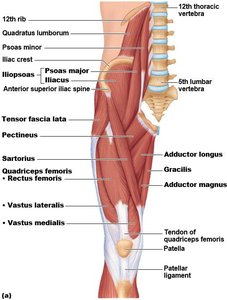
Anterior Thigh Muscles
The anterior thigh muscles are primarily responsible for flexion at the hip and extension at the knee. Key muscles include:
Iliacus: Originates from the iliac fossa, inserts on the lesser trochanter of the femur. Flexes the thigh or trunk.
Psoas major: Originates from lumbar vertebrae, inserts on the lesser trochanter. Flexes vertebral column laterally and flexes thigh or trunk.
Sartorius: The longest muscle in the body, runs obliquely across the anterior thigh. Originates from the anterior superior iliac spine (ASIS), inserts on the tibia. Flexes, abducts, and laterally rotates the thigh; weak knee flexor.
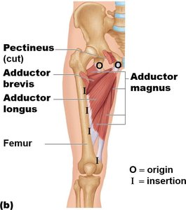
Medial Thigh Muscles
The medial compartment muscles are primarily adductors of the thigh. They include:
Adductor magnus: Originates from the ischium and pubis, inserts on the adductor tubercle of the femur. Adducts the thigh.
Adductor longus: Most anterior adductor, originates from the pubic bone, inserts on the femur. Adducts the thigh.
Adductor brevis: Originates from the pubic bone, inserts on the femur. Adducts the thigh.
Gracilis: Originates from the ischium and pubis, inserts on the tibia. Adducts the thigh.
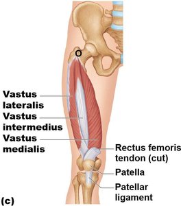
Quadriceps Femoris Group
The quadriceps femoris is a group of four muscles forming the bulk of the anterior thigh, responsible for extension of the leg at the knee. The rectus femoris also flexes the thigh at the hip.
Rectus femoris: Originates from the anterior inferior iliac spine (AIIS), inserts on the tibial tuberosity via the patellar ligament. Extends leg and flexes thigh.
Vastus lateralis: Lateral aspect of thigh, originates from femur, inserts on tibial tuberosity. Extends leg.
Vastus medialis: Inferomedial aspect, originates from femur, inserts on tibial tuberosity. Extends leg.
Vastus intermedius: Deep to rectus femoris, originates from femur, inserts on tibial tuberosity. Extends leg.
Tensor Fascia Lata
The tensor fascia lata is located on the anterolateral aspect of the thigh. It originates from the iliac crest and inserts into the iliotibial tract. Its actions include flexion and abduction of the thigh.
Gluteal Muscles and Lateral Rotators
Gluteal Muscles
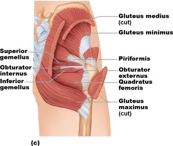
The gluteal muscles are located in the buttock region and are responsible for extension and abduction of the thigh.
Gluteus maximus: Largest and most superficial gluteal muscle, forms the bulk of the buttock. Originates from the ilium, sacrum, and coccyx; inserts on the gluteal tuberosity of the femur. Extends thigh, especially when rising from a flexed position.
Gluteus medius: Covered by gluteus maximus, originates from the ilium, inserts on the greater trochanter. Abducts thigh.
Gluteus minimus: Smallest and deepest gluteal muscle, originates from the ilium, inserts on the greater trochanter. Abducts thigh.
Lateral Rotators of the Hip
Several muscles act to laterally rotate the thigh at the hip joint, including:
Piriformis: Pyramidal muscle on the posterior aspect of the hip joint, originates from the sacrum, inserts on the greater trochanter. Laterally rotates thigh.
Other lateral rotators include the superior and inferior gemellus, obturator internus and externus, and quadratus femoris.
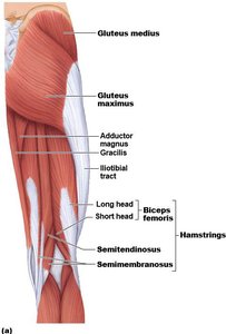
Posterior Thigh Muscles (Hamstrings)
Hamstring Muscles
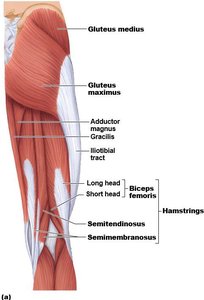
The hamstrings are a group of three muscles located in the posterior compartment of the thigh. They are responsible for extension of the thigh at the hip and flexion of the leg at the knee.
Biceps femoris: Most lateral hamstring, arises from two heads (ischial tuberosity and femur), inserts on the fibula. Extends thigh and flexes leg.
Semitendinosus: Medial to biceps femoris, originates from ischial tuberosity, inserts on tibia. Extends thigh and flexes leg.
Semimembranosus: Deep to semitendinosus, originates from ischial tuberosity, inserts on tibia. Extends thigh and flexes leg.
Muscles of the Leg: Movements of the Ankle and Toes
Leg Compartments and General Actions
The leg is divided into anterior, lateral, and posterior compartments, each containing muscles responsible for specific movements:
Ankle: Dorsiflexion (lifting the foot) and plantarflexion (pointing the foot).
Intertarsal joints: Inversion (turning sole inward) and eversion (turning sole outward).
Toes: Flexion and extension.
Anterior Leg Muscles
These muscles are primarily responsible for dorsiflexion and extension of the toes.
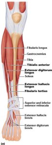
Tibialis anterior: Superficial muscle paralleling the shin, originates from tibia, inserts on medial cuneiform and 1st metatarsal. Dorsiflexion and inversion.
Extensor digitorum longus: Lateral to tibialis anterior, originates from tibia and fibula, inserts on middle and distal phalanges of digits II-V. Extends toes and dorsiflexion.
Extensor hallucis longus: Deep to extensor digitorum longus and tibialis anterior, originates from fibula, inserts on distal phalanx of great toe. Extends great toe and dorsiflexion.
Lateral Leg Muscles
The lateral compartment muscles are involved in plantarflexion and eversion of the foot.
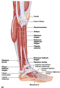
Fibularis longus: Superficial lateral muscle, originates from fibula, inserts on medial cuneiform and 1st metatarsal. Plantarflexion and eversion.
Fibularis brevis: Smaller, deep to fibularis longus, originates from fibula, inserts on 5th metatarsal. Plantarflexion and eversion.
Posterior Leg Muscles
The posterior compartment muscles are responsible for plantarflexion of the foot and flexion of the knee.
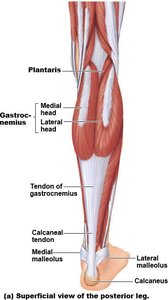
Gastrocnemius: Superficial muscle forming the calf, originates from femoral condyles, inserts on calcaneus. Plantarflexes foot and flexes knee.
Soleus: Broad, flat muscle deep to gastrocnemius, originates from tibia and fibula, inserts on calcaneus. Plantarflexes foot.
Tibialis posterior: Deep to soleus, originates from tibia and fibula, inserts on tarsals and metatarsals II-IV. Inversion and plantarflexion.
Summary Table: Major Muscles Crossing Hip, Knee, and Leg
This table summarizes the origin, insertion, and actions of key muscles discussed above.
Muscle | Origin | Insertion | Action |
|---|---|---|---|
Iliacus | Iliac fossa | Lesser trochanter of femur | Flexes thigh/trunk |
Psoas major | Lumbar vertebrae | Lesser trochanter of femur | Flexes vertebral column laterally; flexes thigh/trunk |
Sartorius | ASIS | Tibia | Flexes, abducts, laterally rotates thigh; weak knee flexor |
Adductor magnus | Ischium and pubis | Adductor tubercle of femur | Adducts thigh |
Rectus femoris | AIIS | Tibial tuberosity via patellar ligament | Extends leg, flexes thigh |
Vastus lateralis/medialis/intermedius | Femur | Tibial tuberosity via patellar ligament | Extends leg |
Gluteus maximus | Ilium, sacrum, coccyx | Gluteal tuberosity of femur | Extends thigh |
Gluteus medius/minimus | Ilium | Greater trochanter of femur | Abducts thigh |
Biceps femoris | Ischial tuberosity, femur | Fibula | Extends thigh, flexes leg |
Semitendinosus/semimembranosus | Ischial tuberosity | Tibia | Extends thigh, flexes leg |
Tibialis anterior | Tibia | Medial cuneiform, 1st metatarsal | Dorsiflexion, inversion |
Fibularis longus/brevis | Fibula | Medial cuneiform/1st metatarsal (longus), 5th metatarsal (brevis) | Plantarflexion, eversion |
Gastrocnemius | Femoral condyles | Calcaneus | Plantarflexes foot, flexes knee |
Soleus | Tibia, fibula | Calcaneus | Plantarflexes foot |
Tibialis posterior | Tibia, fibula | Tarsals, metatarsals II-IV | Inversion, plantarflexion |
Key Terms and Concepts
Origin (O): The fixed attachment point of a muscle.
Insertion (I): The movable attachment point of a muscle.
Flexion: Decreasing the angle between two bones.
Extension: Increasing the angle between two bones.
Abduction: Moving a limb away from the midline.
Adduction: Moving a limb toward the midline.
Dorsiflexion: Lifting the foot upward.
Plantarflexion: Pointing the foot downward.
Inversion: Turning the sole of the foot inward.
Eversion: Turning the sole of the foot outward.
Clinical Relevance
Injuries to the quadriceps or hamstrings can impair walking, running, and jumping.
Weakness in the gluteal muscles may lead to gait abnormalities.
Damage to the tibialis anterior can cause "foot drop," a condition where dorsiflexion is impaired.
Additional info: The anatomical arrangement of these muscles is essential for coordinated movement and stability of the lower limb. Understanding their origins, insertions, and actions is fundamental for clinical assessment and rehabilitation.
