 Indietro
Muscles of the Face, Neck, and Pectoral Girdle: Structure, Function, and Organization
Guida di studio - Note intelligenti
 Appunti personalizzati basati sui tuoi materiali, ampliati con definizioni chiave, esempi e contesto.
Muscles of the Face and Neck
Superficial and Deep Muscles
Muscles of the face and neck are organized by their relative position to the skin and underlying structures. Superficial muscles are closer to the skin, while deep muscles lie beneath other tissues. Understanding these layers is essential for anatomical dissection and clinical practice.
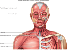
Superficial muscles: Easily visible and often removed to reveal deeper structures. Example: Platysma is superficial to sternocleidomastoid.
Deep muscles: Located beneath superficial muscles, often responsible for more complex movements.
Layer relationship in the neck: Skin → Platysma → Sternocleidomastoid (from superficial to deep).
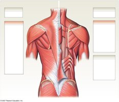
Muscle Groups of the Neck, Shoulder, and Back
The neck, shoulder, and back contain both axial and appendicular muscles, each with distinct functions.
Axial muscles: Support and move the head, neck, and trunk. Examples include sternocleidomastoid, semispinalis capitis, splenius capitis, and erector spinae group.
Appendicular muscles: Move the limbs and stabilize the pectoral girdle. Examples include trapezius, deltoid, infraspinatus, teres minor, teres major, triceps brachii, latissimus dorsi, levator scapulae, supraspinatus, rhomboid minor, rhomboid major, and serratus anterior.

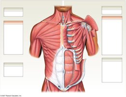
Muscles of the Trunk and Proximal Limb
Superficial and Deep Muscles of the Trunk
The trunk muscles are divided into superficial and deep layers, each contributing to movement and stability.
Superficial muscles: Platysma, deltoid, pectoralis major, latissimus dorsi, serratus anterior, external oblique, rectus sheath, superficial inguinal ring.
Deep muscles: Sternocleidomastoid, trapezius, subclavius, deltoid (cut and reflected), pectoralis minor, subscapularis, coracobrachialis, biceps brachii, teres major, external and internal intercostals, internal oblique, rectus abdominis, transversus abdominis.

Muscles That Position the Pectoral Girdle
Overview and Classification
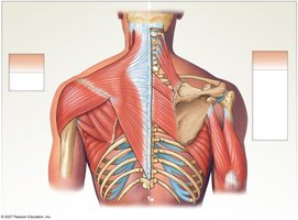
The pectoral girdle is stabilized and moved by several muscles, which are classified as superficial or deep based on their anatomical location.
Superficial muscles: Trapezius, deltoid, infraspinatus, teres minor, teres major, serratus anterior.
Deep muscles: Levator scapulae, rhomboid minor, rhomboid major, serratus anterior, triceps brachii.
Muscles That Position the Pectoral Girdle: Details
Levator scapulae: Elevates scapula and rotates glenoid cavity inferiorly.
Trapezius: Elevates, retracts, and rotates scapula; assists in shoulder movements.
Subclavius: Stabilizes and depresses clavicle.
Pectoralis minor: Stabilizes scapula by drawing it anteriorly and inferiorly.
Serratus anterior: Protracts scapula and holds it against thoracic wall.
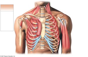



Table: Muscles That Position the Pectoral Girdle
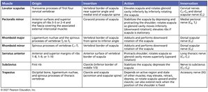
This table summarizes the origin, insertion, action, and innervation of key muscles that position the pectoral girdle.
Muscle | Origin | Insertion | Action | Innervation |
|---|---|---|---|---|
Levator scapulae | Transverse processes of first four cervical vertebrae | Vertebral border of scapula near superior angle | Elevates scapula and rotates glenoid cavity inferiorly | Cervical nerves C3-C4, dorsal scapular nerve (C5) |
Pectoralis minor | Anterior surfaces and superior margins of ribs 3-5 and the fascia covering the associated external intercostal muscles | Coracoid process of scapula | Stabilizes scapula by depressing and rotating it; assists in forced inspiration | Medial pectoral nerve (C8, T1) |
Rhomboid major | Spinous processes of vertebrae T2-T5 | Vertebral border of scapula from spine to inferior angle | Adducts and performs downward rotation of scapula | Dorsal scapular nerve (C5) |
Rhomboid minor | Spinous processes of vertebrae C7-T1 | Vertebral border of scapula near spine | Adducts and performs downward rotation of scapula | Dorsal scapular nerve (C5) |
Serratus anterior | Anterior and superior margins of ribs 1-8, 1-9, or 1-10 | Anterior surface of vertebral border of scapula | Protracts scapula and holds it against thoracic wall | Long thoracic nerve (C5-C7) |
Subclavius | First rib | Clavicle (inferior border) | Stabilizes the scapula; depresses lateral end of clavicle | Nerve to subclavius (C5-C6) |
Trapezius | Occipital bone, ligamentum nuchae, spinous processes of thoracic vertebrae | Clavicle and scapula (acromion and scapular spine) | Depends on active region and state; elevates, retracts, depresses, or rotates scapula | Accessory nerve (CN XI) |
Muscles That Move the Arm
Superficial and Deep Muscles
Muscles that move the arm are classified based on their anatomical location and function. These muscles originate from the pectoral girdle or trunk and insert onto the humerus or forearm.
Superficial muscles: Deltoid, pectoralis major, latissimus dorsi, supraspinatus.
Deep muscles: Subscapularis, coracobrachialis, teres major, biceps brachii, triceps brachii, infraspinatus, teres minor.
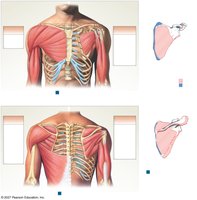
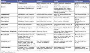
Table: Muscles That Move the Arm
This table summarizes the origin, insertion, action, and innervation of key muscles that move the arm.
Muscle | Origin | Insertion | Action | Innervation |
|---|---|---|---|---|
Coracobrachialis | Coracoid process | Medial margin of shaft of humerus | Assists in adduction and flexion of shoulder | Musculocutaneous nerve (C5-C7) |
Deltoid | Clavicle and scapula (acromion and scapular spine) | Deltoid tuberosity of humerus | Abduction, flexion, extension, and rotation of shoulder | Axillary nerve (C5-C6) |
Supraspinatus | Supraspinous fossa of scapula | Greater tubercle of humerus | Assists the deltoid in abduction of shoulder | Suprascapular nerve (C5) |
Subscapularis | Subscapular fossa of scapula | Lesser tubercle of humerus | Medial rotation of shoulder | Subscapular nerve (C5-C6) |
Teres major | Inferior angle of scapula | Medial lip of intertubercular sulcus of humerus | Extension, adduction, and medial rotation of shoulder | Lower subscapular nerve (C5-C6) |
Biceps brachii (long head) | Supraglenoid tubercle of scapula | Radial tuberosity | Flexion of elbow and shoulder; supination | Musculocutaneous nerve (C5-C6) |
Latissimus dorsi | Spinous processes of inferior thoracic and all lumbar and sacral vertebrae, ribs 8-12, thoracolumbar fascia | Floor of intertubercular sulcus of humerus | Extension, adduction, and medial rotation of shoulder | Thoracodorsal nerve (C6-C8) |
Pectoralis major | Cartilages of ribs 2-6, body of sternum, inferior and medial clavicle | Crest of greater tubercle and lateral lip of intertubercular sulcus of humerus | Flexion, adduction, and medial rotation of shoulder | Medial and lateral pectoral nerves (C5-T1) |
Muscles of the Head and Neck
Functions and Classification
Head and neck muscles are specialized for facial expression, mastication, eye movement, and head/shoulder movement.
Facial expression muscles: Insert into skin or other muscles, allowing a wide range of emotions.
Muscles of mastication: Move the mandible during chewing.
Eye movement muscles: Six extrinsic muscles control eye position.
Neck muscles: Provide movement of the head and shoulder girdle.
Muscles of Facial Expression: Orbital and Nasal Regions
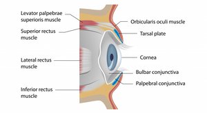
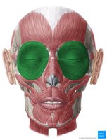
Orbicularis oculi: Circular muscle around the eye; closes eyelids in blinking, squinting, and sleeping; aids tear flow.
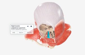
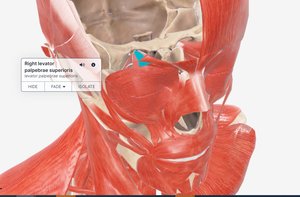
Levator palpebrae superioris: Elevates upper eyelid, opening the eye.
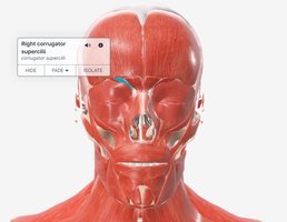
Corrugator supercilii: Draws eyebrows medially and downward; causes forehead wrinkles; reduces glare.
Nasalis: Widens nostrils and narrows internal air passage.
Muscles of Facial Expression: Oral Region
Levator labii superioris: Raises upper lip ("Elvis snarl").
Orbicularis oris: Encircles mouth; closes and protrudes lips ("kissy face").
Zygomaticus major: Raises corners of mouth (smiling/laughing).
Zygomaticus minor: Elevates upper lip, exposes upper teeth.
Risorius: Draws corner of lip laterally; tenses lips in expressions of horror, laughing, or disgust.
Depressor anguli oris: Draws angles of mouth downward (frowning).
Muscles of Facial Expression: Mental & Buccal Regions
Mentalis: Protrudes lower lip and wrinkles chin (pouting).
Buccinator: Draws corners of mouth laterally, compresses cheek against teeth and gums; holds food between teeth during chewing; used in whistling, blowing, or playing instruments.
Muscles of Facial Expression: Scalp Region
Occipitofrontalis (frontal belly): Elevates eyebrows; expressions of surprise or fright; wrinkles forehead.
Muscles of Mastication
Temporalis: Attaches at temporal lines of cranium and coronoid process of mandible; moves mandible up, back, and side to side.
Masseter: Powerful jaw muscle; elevates mandible.
Medial & lateral pterygoid: Assist in grinding movements of the jaw.
Muscles of the Neck
Sternocleidomastoid: Prime mover in neck flexion; attaches from manubrium (sternum) to mastoid process; bilateral contraction flexes neck, unilateral contraction rotates head.
Scalene muscles: Flex and slightly rotate neck.
Muscles of Chewing and Swallowing
Suprahyoid muscles: Digastric (depresses mandible, elevates hyoid), mylohyoid (forms floor of mouth, elevates hyoid and tongue), stylohyoid (elevates hyoid and tongue).
Infrahyoid muscles: Omohyoid (depresses larynx and hyoid), sternohyoid (depresses larynx and hyoid), thyrohyoid (depresses hyoid, elevates larynx), sternothyroid (depresses larynx after elevation).
Neck Regions: The Anterior Triangle
The neck is divided into anatomical regions called "triangles" for clinical and anatomical reference. The anterior triangle is defined by the mandible, sternocleidomastoid, and midline of the neck.
Summary Table: Key Muscles and Their Functions
Superficial muscles: Platysma, sternocleidomastoid, trapezius, deltoid, pectoralis major, latissimus dorsi, serratus anterior, external oblique.
Deep muscles: Semispinalis capitis, splenius capitis, levator scapulae, supraspinatus, rhomboid minor/major, subclavius, subscapularis, coracobrachialis, biceps brachii, teres major/minor, internal intercostals, internal oblique, rectus abdominis, transversus abdominis.
Facial expression muscles: Orbicularis oculi, levator palpebrae superioris, corrugator supercilii, nasalis, levator labii superioris, orbicularis oris, zygomaticus major/minor, risorius, depressor anguli oris, mentalis, buccinator, occipitofrontalis.
Mastication muscles: Temporalis, masseter, medial/lateral pterygoid.
Neck muscles: Sternocleidomastoid, scalene, suprahyoid, infrahyoid.
Additional info: Tables and images included above are inferred to be directly relevant based on their anatomical labeling and context in the notes. All muscle names, actions, and innervations are expanded for academic completeness.
