 Indietro
Muscles of the Head, Neck, and Trunk: Structure, Function, and Identification
Guida di studio - Note intelligenti
 Appunti personalizzati basati sui tuoi materiali, ampliati con definizioni chiave, esempi e contesto.
Muscles of the Head, Neck, and Trunk
Introduction
This study guide covers the anatomy, terminology, and functional organization of the major skeletal muscles of the head, neck, and trunk. It includes muscle naming conventions, mechanisms of muscle action, and the identification and function of key muscle groups. Understanding these muscles is essential for comprehending movement, support, and protection provided by the musculoskeletal system.
Naming Skeletal Muscles
Muscle Naming Conventions
Skeletal muscles are named based on several criteria, including location, position, structure, and function. Recognizing these conventions aids in identifying and understanding muscle roles.
Location: Indicates the region (e.g., abdominis for abdomen, femoris for femur).
Position/Direction: Describes orientation (e.g., anterior, lateralis, rectus for straight, transversus for transverse).
Structural Characteristics: Includes number of origins (e.g., biceps = two heads), shape (e.g., deltoid = triangle), and size (e.g., maximus = largest).
Origin and Insertion: Muscles are often named for their attachment points (e.g., sternocleidomastoid attaches to sternum, clavicle, and mastoid process).
Action: Describes the primary movement (e.g., flexor, extensor).
Example: The rectus abdominis is a straight muscle of the abdomen.
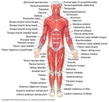
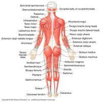
How Skeletal Muscles Produce Movement
Muscle Action and Levers
Skeletal muscles produce movement by exerting force on tendons, which pull on bones or other structures. Most muscles cross at least one joint and are attached to the bones forming that joint. When a muscle contracts, it draws one articulating bone toward the other, creating movement at the joint.
Lever: A rigid object (bone) that moves on a fixed point (fulcrum) when force is applied.
Fulcrum: The fixed point (joint) on which the lever moves.
Tendon: Cordlike structure of dense regular connective tissue connecting muscle to bone.
Aponeurosis: Broad, flat connective tissue sheet connecting muscle to bone or other muscles.
Origin: The stationary attachment point of the muscle.
Insertion: The movable attachment point of the muscle.
Action: The specific movement produced by muscle contraction.
Example: The biceps brachii flexes the forearm at the elbow joint by pulling on the radius.
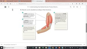
Functional Classification of Muscles
Agonist (Prime Mover): The main muscle responsible for a specific movement.
Antagonist: Muscle that opposes the action of the agonist.
Synergist: Muscle that assists the agonist in performing its action.
Muscles of Facial Expression and Mastication
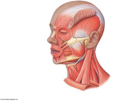
Muscles of Facial Expression
These muscles are responsible for movements of the skin and soft tissues of the face, allowing for expressions such as smiling, frowning, and blinking. They are primarily innervated by the facial nerve (cranial nerve VII).
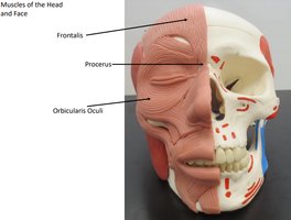
Frontalis: Raises eyebrows and wrinkles forehead.
Orbicularis oculi: Closes eyelids.
Zygomaticus major/minor: Elevates corners of the mouth (smiling).
Orbicularis oris: Closes and protrudes lips (kissing).
Buccinator: Compresses cheek (whistling, blowing).
Platysma: Tenses skin of neck and depresses mandible.
Depressor anguli oris: Depresses corners of the mouth (frowning).
Mentalis: Protrudes lower lip (pouting).
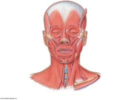
Muscles of Mastication
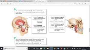
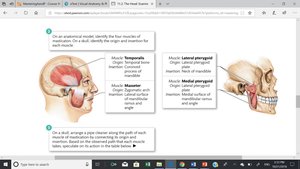
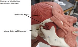
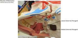
The muscles of mastication are responsible for movements of the jaw during chewing. They act on the temporomandibular joint (TMJ).
Temporalis: Elevates and retracts mandible.
Masseter: Elevates mandible (primary chewing muscle).
Medial pterygoid: Elevates mandible and moves it side to side.
Lateral pterygoid: Depresses and protracts mandible, moves it side to side.
Example: The masseter is the strongest muscle for jaw elevation during chewing.
Model Images of Facial Muscles
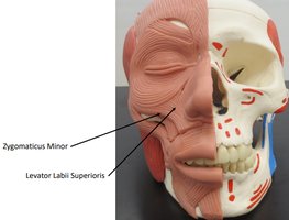
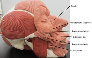
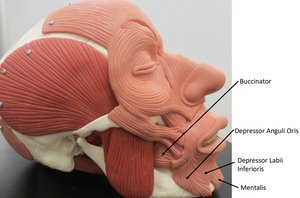
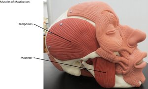
Extrinsic Eye Muscles
Overview
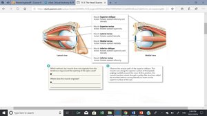
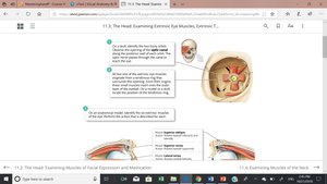
Extrinsic eye muscles control the movement of the eyeball within the orbit. Each muscle has a specific origin, insertion, and action, allowing for precise eye movements.
Superior rectus: Elevates and turns eye medially.
Inferior rectus: Depresses and turns eye medially.
Lateral rectus: Moves eye laterally.
Medial rectus: Moves eye medially.
Superior oblique: Depresses and turns eye laterally.
Inferior oblique: Elevates and turns eye laterally.
Muscles of the Neck
Major Muscles and Triangles of the Neck
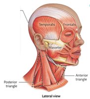
The neck contains several important muscles that facilitate head movement, swallowing, and support of the hyoid bone. The neck is divided into anatomical triangles for clinical reference.
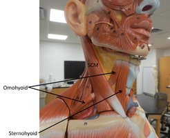
Sternocleidomastoid (SCM): Flexes and rotates the head.
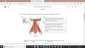
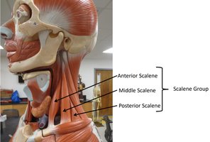
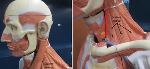
Scalene muscles (anterior, middle, posterior): Elevate first two ribs, assist in neck flexion and rotation.
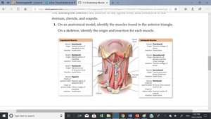
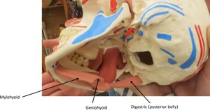
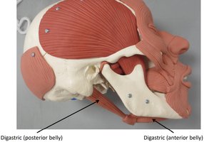
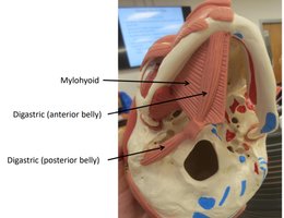
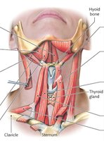
Suprahyoid muscles (geniohyoid, mylohyoid, stylohyoid, digastric): Elevate hyoid bone, assist in swallowing.
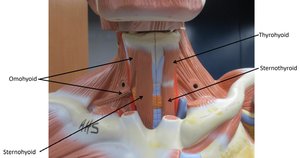
Infrahyoid muscles (sternohyoid, omohyoid, sternothyroid, thyrohyoid): Depress hyoid bone and larynx during swallowing and speech.
Muscles of the Thorax
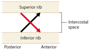
Intercostal Muscles
The intercostal muscles are located between the ribs and are essential for respiration.
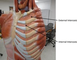
External intercostals: Elevate ribs during inspiration.
Internal intercostals: Depress ribs during forced expiration.
Diaphragm
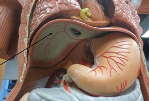
The diaphragm is the primary muscle of respiration, separating the thoracic and abdominopelvic cavities.
Origin: Xiphoid process, costal cartilages, lumbar vertebrae.
Insertion: Central tendon of the diaphragm.
Action: Contracts to increase thoracic volume during inspiration.
Pectoral Muscles
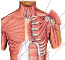
Pectoralis major: Flexes, adducts, and medially rotates the arm.
Pectoralis minor: Depresses and protracts the scapula.
Muscles of the Abdominal Wall
Major Muscles and Structures
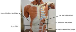
The abdominal wall is composed of several layers of muscle that protect abdominal organs, assist in trunk movement, and maintain intra-abdominal pressure.
Rectus abdominis: Flexes vertebral column, compresses abdomen.
External oblique: Compresses abdomen, flexes and rotates vertebral column.
Internal oblique: Compresses abdomen, flexes and rotates vertebral column.
Transversus abdominis: Compresses abdomen.
Linea alba: Midline tendinous seam joining the abdominal muscles.
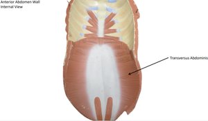
Summary Table: Key Muscles of the Head, Neck, and Trunk
Muscle | Origin | Insertion | Action |
|---|---|---|---|
Temporalis | Temporal bone | Coronoid process of mandible | Elevates and retracts mandible |
Masseter | Zygomatic arch | Mandibular ramus | Elevates mandible |
Sternocleidomastoid | Sternum and clavicle | Mastoid process of temporal bone | Flexes and rotates head |
Rectus abdominis | Pubic crest and symphysis | Costal cartilages of ribs 5-7, xiphoid process | Flexes vertebral column, compresses abdomen |
External intercostals | Inferior border of rib above | Superior border of rib below | Elevates ribs during inspiration |
Diaphragm | Xiphoid process, lower ribs, lumbar vertebrae | Central tendon | Increases thoracic cavity volume |
Key Terms and Concepts
Origin: Fixed attachment point of a muscle.
Insertion: Movable attachment point of a muscle.
Action: Movement produced by muscle contraction.
Agonist, Antagonist, Synergist: Functional roles in movement.
Tendon, Aponeurosis: Connective tissue structures for muscle attachment.
Additional info: For exam preparation, students should be able to identify the origin, insertion, and action of each muscle listed, as well as recognize these muscles on anatomical models and diagrams.
