 Indietro
Muscles of the Head, Neck, Trunk, and Pelvic Floor: Structure and Function
Guida di studio - Note intelligenti
 Appunti personalizzati basati sui tuoi materiali, ampliati con definizioni chiave, esempi e contesto.
Muscles of Facial Expression
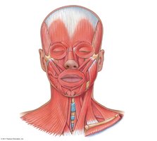
Overview of Facial Muscles
Muscles of facial expression are unique in that they do not move joints but instead manipulate the skin to produce expressions. These muscles are essential for nonverbal communication and are innervated primarily by the facial nerve (cranial nerve VII).
Corrugator supercilii: Pulls eyebrows together, creating a frown.
Levator labii superioris: Elevates upper lip.
Levator anguli oris: Elevates angle of mouth.
Depressor anguli oris: Depresses angle of mouth.
Depressor labii inferioris: Depresses lower lip.
Mentalis: Wrinkles skin on the chin, produces a pout.
Orbicularis oculi: Closes eyes.
Zygomaticus minor: Elevates upper lip.
Zygomaticus major: Elevates corner of upper lip.
Risorius: Pulls corner of mouth posteriorly.
Frontalis: Raises eyebrows.
Orbicularis oris: Puckers lips.
Muscles of Mastication (Chewing)
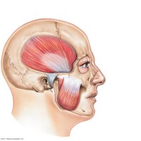
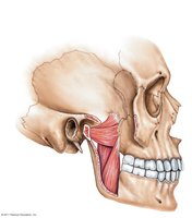
The muscles of mastication are responsible for movements of the jaw, allowing for chewing and speaking. These muscles act on the temporomandibular joint (TMJ).
Masseter: Elevates mandible, primary muscle for jaw closure.
Temporalis: Elevates mandible, assists in jaw retraction.
Buccinator: Holds cheek to the teeth, assists in chewing.
Medial and Lateral Pterygoid: Move the jaw side-to-side and forward (protraction).
Muscles of the Tongue
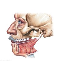
The tongue muscles are crucial for speech, swallowing, and manipulating food.
Palatoglossus: Elevates posterior part of tongue.
Styloglossus: Retracts and elevates tongue.
Genioglossus: Protrudes and depresses tongue.
Hyoglossus: Depresses and retracts tongue.
Muscles of the Neck
Anterior Neck Muscles
The anterior neck muscles are involved in swallowing, speech, and movement of the head.
Mylohyoid, Digastric, Geniohyoid: Muscles of the floor of the mouth, assist in swallowing.
Omohyoid, Sternohyoid, Thyrohyoid, Sternothyroid: Small muscles associated with the hyoid bone, involved in swallowing and speech.
Sternocleidomastoid: Major muscle for neck flexion and rotation; contralateral rotation of neck.
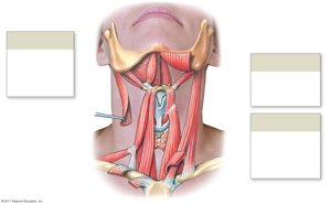
Movements: Ipsilateral vs. Contralateral Rotation
Ipsilateral rotation: Muscle causes movement to the same side as the muscle's location.
Contralateral rotation: Muscle causes movement to the opposite side.
Muscles Moving the Head and Trunk
Sternocleidomastoid: Contralateral rotation and lateral flexion of neck; elevates ribs during deep inhalation.
Scalenes (anterior, middle, posterior): Lateral flexion of neck, elevate ribs.
Splenius capitis and cervicis: Extend neck, ipsilateral rotation, lateral flexion.
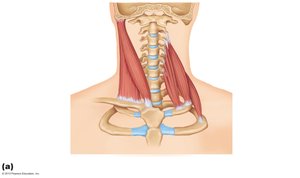
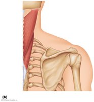
Muscles of the Vertebral Column
Erector Spinae and Deep Back Muscles
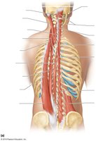
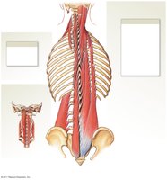
The erector spinae group is the primary extensor of the vertebral column, maintaining posture and allowing trunk movement.
Erector spinae: Includes spinalis, longissimus, and iliocostalis muscle groups; extends trunk and neck.
Semispinalis group: Deep extensors of the spine.
Quadratus lumborum: Lateral flexion of trunk, pelvis elevation.
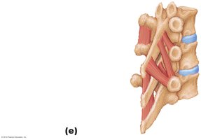
Multifidus, Rotatores, Interspinales, Intertransversarius: Intrinsic muscles of the spine, contribute to movement and proprioception.
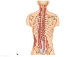
Muscles of Respiration
Intercostal Muscles
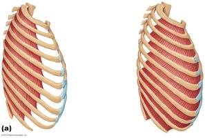
The intercostal muscles are essential for breathing by moving the rib cage.
External intercostal: Elevate ribs during inhalation.
Internal intercostal: Depress ribs during exhalation.
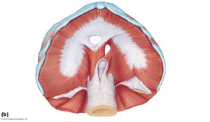
Diaphragm
The diaphragm is the primary muscle of respiration, separating the thoracic and abdominal cavities.
Diaphragm: Contracts to increase thoracic volume, causing inhalation.
Foramen for esophagus, inferior vena cava, and aorta: Openings for major structures passing through the diaphragm.
Muscles of the Abdominal Wall
Abdominal Muscles and Their Functions
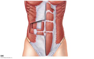
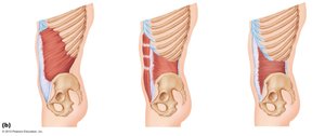
The abdominal wall muscles support trunk movement, protect internal organs, and compress the abdominal cavity.
Rectus abdominis: Flexes trunk.
External oblique: Flexes trunk, laterally flexes trunk, contralateral rotation.
Internal oblique: Flexes trunk, laterally flexes trunk, ipsilateral rotation.
Transversus abdominis: Compresses abdominal cavity.
Serratus anterior: Assists in movement of the scapula.
Muscles of the Pelvic Floor and Perineum
Pelvic Diaphragm and Associated Muscles
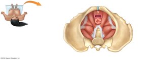
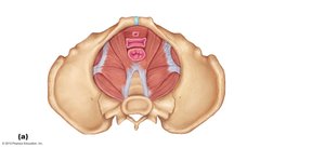
The pelvic floor muscles support pelvic organs, control openings of the urethra, vagina, and anus, and assist in maintaining continence.
Levator ani: Includes pubococcygeus and iliococcygeus; supports pelvic viscera.
Coccygeus: Supports pelvic floor.
Urogenital diaphragm: Contains muscles that control the urethra and vagina.
Piriformis: Assists in lateral rotation of hip.
Summary Table: Major Muscle Groups and Functions
Muscle Group | Main Function |
|---|---|
Facial Expression | Move skin for expressions |
Mastication | Chewing, jaw movement |
Tongue | Speech, swallowing |
Neck | Head movement, swallowing |
Back (Erector Spinae) | Trunk/neck extension |
Intercostals | Breathing |
Diaphragm | Breathing |
Abdominal Wall | Trunk movement, compression |
Pelvic Floor | Support pelvic organs |
Additional info: All four abdominal muscles compress the abdominal cavity, contributing to core stability and protection of internal organs.
