 Indietro
Muscular System: Histology and Physiology Study Guide
Guida di studio - Note intelligenti
 Appunti personalizzati basati sui tuoi materiali, ampliati con definizioni chiave, esempi e contesto.
Muscular System: Histology and Physiology
Functions and Properties of Muscle Tissue
The muscular system is essential for movement, posture, and various physiological functions. Muscle tissue possesses unique properties that enable its function.
Movement of the body: Muscles contract to produce movement.
Maintenance of posture: Continuous muscle activity stabilizes body position.
Respiration: Muscles such as the diaphragm facilitate breathing.
Production of body heat: Muscle contractions generate heat, maintaining body temperature.
Communication: Muscles enable facial expressions, speech, and gestures.
Constriction of organs and vessels: Smooth muscle regulates internal organ function.
Contraction of the heart: Cardiac muscle pumps blood throughout the body.
Stabilize joints: Muscles reinforce and stabilize joints.
Contractility: Ability to shorten forcefully.
Excitability: Ability to respond to stimuli.
Conductivity: Ability to propagate electrical signals.
Extensibility: Ability to stretch without damage.
Elasticity: Ability to return to original shape after stretching.
Types of Muscle Tissue
Muscle tissue is classified into three types: skeletal, cardiac, and smooth. Each type has distinct structural and functional characteristics.
Type of Muscle Tissue | Structure | Location | Voluntary/Involuntary | Function |
|---|---|---|---|---|
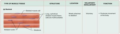
Skeletal | Long, cylindrical, striated fibers; multinucleated | Mostly attached to skeleton | Voluntary | Produces movement of the body |
Skeletal Muscle Tissue Structure
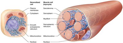
Skeletal muscle fibers are specialized cells with unique structures that facilitate contraction.
Endomysium: Connective tissue surrounding each muscle fiber.
Sarcolemma: Plasma membrane of the muscle cell.
Sarcoplasm: Cytoplasm of the muscle cell, rich in glycogen and myoglobin.
Sarcoplasmic reticulum (SR): Specialized endoplasmic reticulum for calcium storage.
Myofibrils: Bundles of contractile proteins within the cell.

Organization of Skeletal Muscle
Skeletal muscle fibers are organized into fascicles, which are grouped to form muscles. Connective tissue layers provide structure and transmit force.
Fascicle: Bundle of muscle fibers surrounded by perimysium.
Epimysium: Connective tissue surrounding the entire muscle.
Tendon: Connective tissue joining muscle to bone.
Fascia: Thick connective tissue enclosing muscles.
Muscle Fiber Structure and Myofibrils
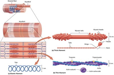
Muscle fibers contain myofibrils, which are composed of myofilaments. These filaments are responsible for muscle contraction.
Thick filaments: Composed of myosin.
Thin filaments: Composed of actin, tropomyosin, and troponin.
Elastic filaments: Composed of titin, providing elasticity and stability.
Sarcomere Structure
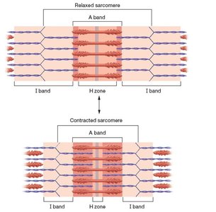
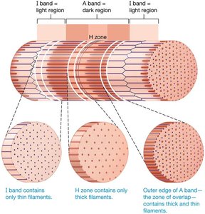
The sarcomere is the functional unit of muscle contraction, defined by specific regions:
I band: Contains only thin filaments.
Z disc: Boundary of the sarcomere; anchors thin filaments.
A band: Contains both thick and thin filaments.
H zone: Contains only thick filaments.
M line: Center of the sarcomere; anchors thick filaments.
Sliding Filament Mechanism of Contraction
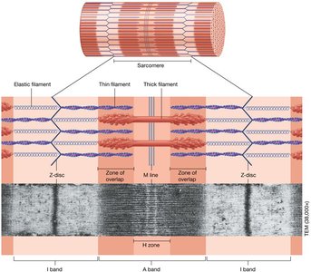
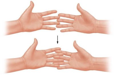
Muscle contraction occurs via the sliding filament mechanism, where myosin heads pull actin filaments toward the M line, shortening the sarcomere.
Myosin heads: Bind to actin, forming cross-bridges.
ATP: Required for myosin head detachment and reactivation.
Dystrophin and Muscular Disease
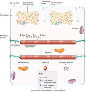
Dystrophin is a structural protein linking actin to the sarcolemma and endomysium. Its absence leads to degenerative muscular diseases, such as Duchenne muscular dystrophy.
Dystrophin: Transfers force of contraction to connective tissue.
Degenerative muscular disease: Most commonly affects boys.
Membrane Potentials in Muscle Cells
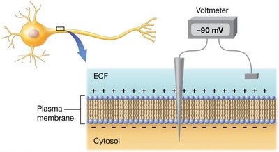
Muscle cells maintain a resting membrane potential due to unequal ion distribution, creating an electrical gradient.
Resting membrane potential: Electrical potential across the plasma membrane.
Voltage: Difference in charge between two points.
Electrical gradient: Source of potential energy.
Ion Channels and Gradients
Ion channels regulate the movement of ions across the plasma membrane, essential for electrical changes and muscle contraction.
Leak channels: Allow passive ion movement.
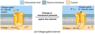
Gated channels: Open in response to specific stimuli.
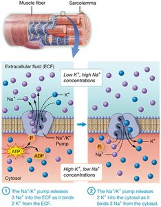
Na+/K+ pump: Maintains concentration gradients by moving 3 Na+ out and 2 K+ in per ATP hydrolyzed.
Electrochemical Gradients
The movement of ions across membranes is determined by both concentration and electrical gradients, collectively termed the electrochemical gradient.
Concentration gradient: Drives diffusion of ions.
Electrical gradient: Attracts ions to oppositely charged regions.
Electrochemical gradient: Combined effect of both gradients.
Generation of the Resting Membrane Potential
To achieve the resting state, the membrane potential must become more negative, requiring the cell to lose more positive charges than it gains.
Positive ions: Their movement creates a negative resting membrane potential.
Action Potentials
An action potential is a rapid change in membrane potential, essential for muscle contraction.
Depolarization: Voltage-gated Na+ channels open; Na+ flows inward.
Repolarization: Na+ channels close; voltage-gated K+ channels open, allowing K+ to diffuse out.
Neuromuscular Junction (NMJ)
The NMJ is the synapse where a motor neuron communicates with muscle fibers, triggering contraction.
Axon terminal/synaptic knob: Releases neurotransmitter.
Synaptic cleft: Space between neuron and muscle fiber.
Motor end plate: Region of sarcolemma with receptors for neurotransmitter.
Phases of Skeletal Muscle Contraction and Relaxation
Muscle contraction and relaxation occur in four major phases:
Excitation: Nerve action potentials lead to muscle action potentials.
Excitation–contraction coupling: Links action potentials to activation of myofilaments.
Contraction: Muscle fiber develops tension and may shorten.
Relaxation: Muscle fiber returns to resting length.
Crossbridge Cycle and Sliding Filament Mechanism
The crossbridge cycle describes the interaction between myosin and actin during contraction.
Myosin heads: Attach, pull, detach, and re-cock, causing filaments to slide toward the M line.
ATP: Required for detachment and reactivation of myosin heads.
Skeletal Muscle Relaxation
Relaxation occurs when calcium is pumped back into the SR, and myosin heads detach from actin.
Energy for Muscle Contraction
ATP is required for multiple processes in muscle contraction and relaxation.
Na+/K+ pumps: Maintain ion gradients.
Myosin head release: Detachment from actin.
Calcium pump: Returns Ca++ to SR.
ATP production: Immediate (creatine phosphate), glycolytic (cytosol), oxidative (mitochondria).
Twitch Contraction and Tension Production
A twitch contraction consists of three phases: latent, contraction, and relaxation. Tension varies with timing, frequency, fiber length, and type.
Wave summation: Increased tension from repeated stimulation.
Tetanus: Unfused and fused forms.
Length-tension relationship: Optimal sarcomere length generates maximal tension.
Motor Units and Recruitment
A motor unit consists of a motor neuron and all muscle fibers it innervates. Recruitment increases force by activating more motor units.
Muscle tone: Baseline involuntary activation.
Types of Muscle Contractions
Muscle contractions are classified by changes in tension and length.
Isotonic contractions: Tension constant, length changes.
Isometric contractions: Length unchanged, tension varies.
Physical Training and Muscle Adaptation
Muscle structure changes in response to training (myoplasticity). Satellite cells aid repair; changes occur within fibers, not in fiber number.
Atrophy: Decrease in muscle size.
Hypertrophy: Increase in muscle size.
Muscular Fatigue and Recovery
Fatigue is the inability to maintain intensity, caused by metabolite depletion, oxygen shortage, chemical accumulation, and environmental factors. Recovery involves increased oxygen consumption (EPOC).
Smooth Muscle
Smooth muscle cells lack striations and motor end plates, and have less extensive SR. They contract via a cascade involving calmodulin and myosin light chain kinase.
Single unit: Cells contract together.
Multi-unit: Cells contract independently.
Cardiac Muscle
Cardiac muscle cells are shorter, branched, and have one or two nuclei. They contain abundant myoglobin and mitochondria, and are linked by intercalated discs (gap junctions and desmosomes). Contraction is involuntary and coordinated by pacemaker cells.
Additional info: Academic context and explanations have been expanded for clarity and completeness. Only images directly relevant to the adjacent content have been included.
