 Indietro
Muscular System: Structure, Function, and Key Muscles of the Human Body
Guida di studio - Note intelligenti
 Appunti personalizzati basati sui tuoi materiali, ampliati con definizioni chiave, esempi e contesto.
Muscular System Overview
Introduction to Muscle Anatomy
The muscular system is essential for movement, stability, and various physiological functions. Muscles are classified by their structure, location, and function, and are organized into groups that work together to produce coordinated actions.
Muscle Types: Skeletal, cardiac, and smooth muscles, with skeletal muscles being the primary focus in anatomy and physiology.
Functions: Movement, posture maintenance, joint stabilization, and heat generation.
Muscle Shapes and Arrangements
Classification of Skeletal Muscle Shapes
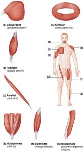
Skeletal muscles are categorized based on their fascicle arrangement, which influences their function and strength.
Circular: Fascicles arranged in concentric rings; found in sphincters (e.g., orbicularis oris).
Convergent: Broad origin, fascicles converge toward a single tendon (e.g., pectoralis major).
Parallel: Fascicles run parallel to the long axis (e.g., sartorius).
Fusiform: Spindle-shaped with a central belly (e.g., biceps brachii).
Multipennate: Fascicles attach obliquely to multiple tendons (e.g., deltoid).
Bipennate: Fascicles insert into the tendon from both sides (e.g., rectus femoris).
Unipennate: Fascicles insert into one side of the tendon (e.g., extensor digitorum longus).
Major Muscles of the Human Body
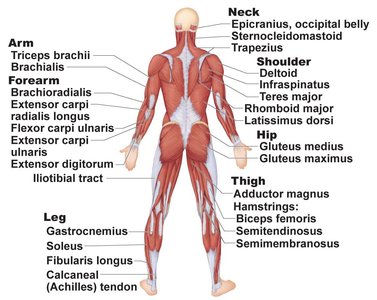
Posterior View: Key Muscles and Their Locations
Understanding the location and function of major muscles is crucial for identifying their roles in movement and posture.
Neck: Includes epicranius, sternocleidomastoid, and trapezius.
Shoulder: deltoid, infraspinatus, teres major, rhomboid major, latissimus dorsi.
Arm: triceps brachii.
Forearm: brachioradialis, extensor carpi radialis longus, flexor carpi ulnaris, extensor digitorum.
Hip: gluteus medius, gluteus maximus.
Thigh: adductor magnus, hamstrings (biceps femoris, semitendinosus, semimembranosus).
Leg: gastrocnemius, soleus, fibularis longus, calcaneal (Achilles) tendon.
Muscles of the Head and Neck
Facial Muscles and Their Functions
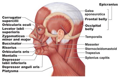
The muscles of the head and neck are responsible for facial expressions, mastication, and movement of the head.
Epicranius: Composed of the frontal and occipital bellies connected by the galea aponeurotica; raises eyebrows and moves scalp.
Orbicularis oculi: Closes the eyelids.
Orbicularis oris: Closes and protrudes lips.
Buccinator: Compresses cheek; important in chewing.
Zygomaticus: Raises corners of mouth (smiling).
Masseter and temporalis: Main muscles of mastication.
Platysma: Tenses skin of neck.
Mastication Muscles
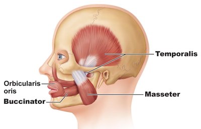
Mastication involves several muscles that move the jaw for chewing.
Masseter: Elevates mandible (jaw closure).
Temporalis: Elevates and retracts mandible.
Buccinator: Assists in mastication by keeping food between teeth.
Pterygoid Muscles
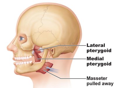
The pterygoid muscles assist in jaw movements, including side-to-side motion.
Lateral pterygoid: Protracts mandible and moves it side-to-side.
Medial pterygoid: Elevates mandible and moves it side-to-side.
Muscles of the Anterior Neck
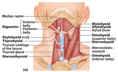
These muscles are involved in swallowing and movement of the hyoid bone and larynx.
Digastric: Has anterior and posterior bellies; depresses mandible, elevates hyoid.
Mylohyoid, stylohyoid, omohyoid, sternohyoid, thyrohyoid: Elevate or depress hyoid bone and larynx.
Sternocleidomastoid: Flexes and rotates the head.
Muscles of the Pharynx and Swallowing
Pharyngeal Constrictors and Associated Muscles
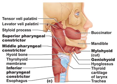
The pharyngeal constrictors are essential for swallowing, moving food from the mouth to the esophagus.
Superior, middle, and inferior pharyngeal constrictors: Sequentially constrict the pharynx during swallowing.
Mylohyoid, geniohyoid, hyoglossus: Assist in tongue and hyoid movements.
Muscles of the Eye
Extraocular Muscles and Their Actions
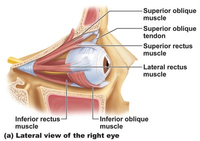
The extraocular muscles control eye movement and are innervated by specific cranial nerves.
Superior rectus: Elevates eye and turns it medially.
Inferior rectus: Depresses eye and turns it medially.
Lateral rectus: Moves eye laterally.
Medial rectus: Moves eye medially.
Superior oblique: Depresses eye and turns it laterally.
Inferior oblique: Elevates eye and turns it laterally.
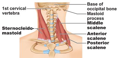
Summary Table: Eye Muscles, Actions, and Cranial Nerves
Muscle | Action | Controlling cranial nerve |
|---|---|---|
Lateral rectus | Moves eye laterally | VI (abducens) |
Medial rectus | Moves eye medially | III (oculomotor) |
Superior rectus | Elevates eye and turns it medially | III (oculomotor) |
Inferior rectus | Depresses eye and turns it medially | III (oculomotor) |
Inferior oblique | Elevates eye and turns it laterally | III (oculomotor) |
Superior oblique | Depresses eye and turns it laterally | IV (trochlear) |
Muscles of the Neck and Back
Neck Muscles: Scalene and Sternocleidomastoid
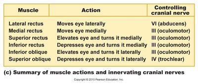
The scalene muscles and sternocleidomastoid are important for head movement and breathing.
Sternocleidomastoid: Flexes and rotates the head.
Scalene muscles (anterior, middle, posterior): Elevate first two ribs, aid in neck flexion.
Back Muscles: Splenius and Erector Spinae
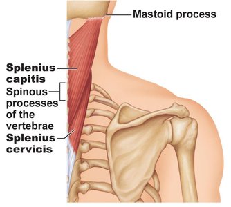
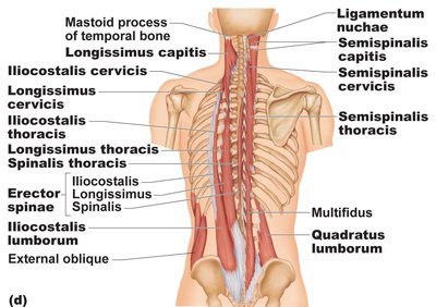
The splenius and erector spinae groups are responsible for extension, rotation, and lateral flexion of the head and vertebral column.
Splenius capitis and cervicis: Extend and rotate the head.
Erector spinae (iliocostalis, longissimus, spinalis): Extend and laterally flex the vertebral column.
Semispinalis, multifidus, quadratus lumborum: Stabilize and move the spine.
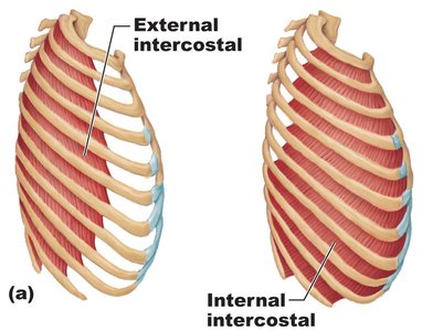
Muscles of the Thorax: Breathing
Intercostal Muscles
The intercostal muscles are located between the ribs and are essential for respiration.
External intercostals: Elevate ribs during inspiration.
Internal intercostals: Depress ribs during forced expiration.
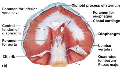
Diaphragm
The diaphragm is the primary muscle of inspiration, separating the thoracic and abdominal cavities.
Diaphragm: Contracts to increase thoracic volume, drawing air into the lungs.
Central tendon: The insertion point for muscle fibers.
Foramina: Openings for the inferior vena cava, esophagus, and aorta.
Summary
Key Points for Exam Preparation
Muscle shape and arrangement affect function and strength.
Major muscles are organized by region and function.
Muscles of the head, neck, back, thorax, and eye have specialized roles in movement, expression, and physiological processes.
Understanding innervation and actions is crucial for clinical and academic applications.
