 Indietro
Muscular Tissue: Structure, Function, and Physiology
Guida di studio - Note intelligenti
 Appunti personalizzati basati sui tuoi materiali, ampliati con definizioni chiave, esempi e contesto.
Muscular Tissue
Overview of Muscle Tissue
Muscular tissue is essential for producing movement, stabilizing body positions, storing and mobilizing substances, and generating heat. It comprises nearly half of the human body’s mass and is specialized for converting chemical energy (ATP) into mechanical energy.
Three Types of Muscle Tissue: Skeletal, Cardiac, and Smooth
Muscle Fiber: Only skeletal and smooth muscle cells are elongated and referred to as muscle fibers.
Main Characteristics: Excitability, contractility, extensibility, and elasticity.
Functions of Muscle Tissue
Producing body movements
Stabilizing body positions
Storing and mobilizing substances
Generating heat
Skeletal Muscle Structure and Organization
Levels of Organization
Skeletal muscle is organized into several hierarchical levels, each with distinct structural and functional properties.
Skeletal muscle (whole muscle)
Muscle fascicle (bundle of fibers)
Muscle fiber (muscle cell)
Myofibril (contractile organelle)
Myofilaments (thick and thin filaments)
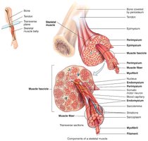
Connective Tissue Sheaths
Each skeletal muscle and muscle fiber is covered by connective tissue sheaths that support and reinforce the muscle:
Epimysium: Surrounds the entire muscle
Perimysium: Surrounds fascicles (bundles of muscle fibers)
Endomysium: Surrounds individual muscle fibers
Microscopic Anatomy of Skeletal Muscle Fibers
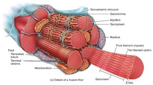
Skeletal muscle fibers are multinucleated, striated cells containing specialized organelles for contraction.
Sarcolemma: Plasma membrane of the muscle fiber
Sarcoplasm: Cytoplasm containing glycosomes (glycogen storage) and myoglobin (O2 storage)
Myofibrils: Densely packed, rodlike elements responsible for muscle contraction
Sarcoplasmic reticulum (SR): Stores and releases Ca2+
Transverse (T) tubules: Conduct action potentials into the muscle fiber
Muscle Fiber Formation and Regeneration
Development and Growth
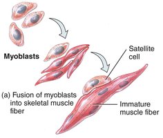
Skeletal muscle fibers are formed by the fusion of myoblasts during embryonic development, resulting in multinucleated cells. Mature muscle fibers cannot divide, but satellite cells (a type of stem cell) can assist in repair and limited regeneration.
Hypertrophy: Enlargement of existing muscle fibers due to increased organelles and proteins
Atrophy: Decrease in muscle fiber size due to loss of myofibrils (aging or disuse)
Fibrosis: Replacement of muscle tissue with fibrous scar tissue if regeneration is insufficient
Myofibrils and Sarcomeres
Structure of Myofibrils
Myofibrils are composed of repeating units called sarcomeres, which are the functional contractile units of muscle fibers. Sarcomeres are defined by Z discs and contain thick (myosin) and thin (actin) filaments arranged in a precise pattern.
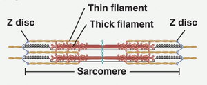
A band: Dark region containing thick filaments
I band: Light region containing thin filaments
H zone: Lighter region in the middle of the A band
M line: Line of proteins that bisects the H zone
Z disc: Boundary of each sarcomere
Molecular Composition of Myofilaments
Thick filaments: Composed of myosin molecules with heads that bind ATP and actin
Thin filaments: Composed of actin, tropomyosin, and troponin
Cross-bridges: Formed when myosin heads bind to actin during contraction
Neuromuscular Junction (NMJ) and Muscle Excitation
Structure and Function of the NMJ
The neuromuscular junction is the site where a motor neuron communicates with a skeletal muscle fiber to initiate contraction. It consists of the axon terminal, synaptic cleft, and motor end plate.
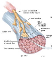
Axon terminal: Releases acetylcholine (ACh) into the synaptic cleft
Motor end plate: Region of sarcolemma with ACh receptors
Synaptic cleft: Space between neuron and muscle fiber
Events at the NMJ
Action potential arrives at axon terminal
Voltage-gated Ca2+ channels open, triggering ACh release
ACh binds to receptors on the motor end plate, opening Na+ channels
Muscle action potential is generated
Acetylcholinesterase degrades ACh, ending the signal
Muscle Contraction: Sliding Filament Mechanism
Steps of the Contraction Cycle
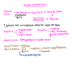
Muscle contraction occurs through the sliding filament mechanism, where thin filaments slide past thick filaments, shortening the sarcomere without changing the length of the filaments themselves.
ATP Hydrolysis: Myosin head hydrolyzes ATP, becoming energized
Cross-Bridge Formation: Myosin head binds to actin
Power Stroke: Myosin head pivots, pulling actin toward the center of the sarcomere
Detachment: New ATP binds to myosin, causing it to detach from actin
Excitation-Contraction Coupling
This process links the muscle action potential to the sliding of filaments and contraction. The action potential travels along the sarcolemma and T tubules, triggering Ca2+ release from the SR, which initiates contraction.
Types of Muscle Contractions
Isotonic vs. Isometric Contractions
Isotonic: Muscle changes length while tension remains constant (movement occurs)
Isometric: Muscle tension increases without changing length (no movement)
Control of Muscle Tension
Motor Units and Recruitment
A motor unit consists of a motor neuron and all the muscle fibers it innervates. The strength of contraction depends on the number of active motor units (motor unit recruitment).
Muscle Twitch: Brief contraction in response to a single action potential
Summation: Increased frequency of stimulation leads to stronger contractions
Tetanus: Sustained contraction with no relaxation between stimuli
Muscle Metabolism and Fatigue
ATP Production Pathways
Creatine Phosphate (CP): Rapid ATP generation for short bursts
Anaerobic Glycolysis: Produces ATP quickly without oxygen, yields lactic acid
Aerobic Respiration: Generates most ATP using oxygen, slower but efficient
Muscle Fatigue
Fatigue is the inability to maintain force of contraction due to ionic imbalances, depletion of energy stores, or accumulation of metabolic byproducts.
Types of Skeletal Muscle Fibers
Classification and Function
Type I (Slow-twitch): High fatigue resistance, aerobic metabolism, endurance activities
Type IIa (Fast-twitch, intermediate): Combination of power and endurance
Type IIb (Fast-twitch): High power, low endurance, anaerobic metabolism
Cardiac and Smooth Muscle
Cardiac Muscle
Found only in the heart
Striated, involuntary, contains intercalated discs for synchronized contraction
High mitochondrial content, long contraction duration
Smooth Muscle
Found in walls of hollow organs (e.g., stomach, blood vessels)
Non-striated, involuntary, can stretch and contract more than other muscle types
Greater regenerative capacity compared to skeletal and cardiac muscle
Regeneration of Muscle Tissue
Skeletal muscle: Limited regeneration via satellite cells
Cardiac muscle: Minimal regeneration
Smooth muscle: Can regenerate from pericytes
Summary Table: Major Features of Muscle Tissue Types
Feature | Skeletal Muscle | Cardiac Muscle | Smooth Muscle |
|---|---|---|---|
Control | Voluntary | Involuntary | Involuntary |
Striations | Yes | Yes | No |
Regeneration | Limited | Very limited | High |
Location | Attached to bones | Heart | Walls of hollow organs |
Key Terms and Concepts
Excitability: Ability to respond to stimuli
Contractility: Ability to shorten forcibly
Extensibility: Ability to be stretched
Elasticity: Ability to recoil to resting length
Cross-bridge: Connection formed between myosin head and actin during contraction
Motor unit: Motor neuron and all muscle fibers it innervates
