 Indietro
Nerve Impulses and Synaptic Transmission: Cells, Action Potentials, and Synapses
Guida di studio - Note intelligenti
 Appunti personalizzati basati sui tuoi materiali, ampliati con definizioni chiave, esempi e contesto.
Cells of the Nervous System
Neuroglia (Glial Cells) of the CNS
Nervous tissue is composed of two principal cell types: neuroglia and neurons. Neuroglia are supportive cells that maintain the environment for neurons, while neurons are excitable cells responsible for transmitting electrical signals.
Astrocytes: Most abundant glial cells; support neurons, regulate exchanges between capillaries and neurons, guide neuron migration, control the chemical environment, respond to nerve impulses, and participate in information processing.
Microglial cells: Small, ovoid cells that monitor neuron health, migrate toward injured neurons, and can phagocytize microorganisms and debris.
Ependymal cells: Line the central cavities of the brain and spinal cord; may be ciliated to circulate cerebrospinal fluid (CSF); form a barrier between CSF and tissue fluid.
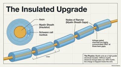
Oligodendrocytes: Branched cells that wrap CNS nerve fibers, forming insulating myelin sheaths.
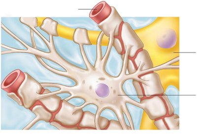
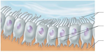
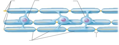
Neuroglia of the PNS
Satellite cells: Surround neuron cell bodies in the PNS; function similarly to astrocytes.
Schwann cells: Surround all peripheral nerve fibers and form myelin sheaths; vital for regeneration of damaged peripheral nerve fibers.
Neurons (Nerve Cells)
Neurons are the structural and functional units of the nervous system. They are highly specialized for impulse conduction and possess unique characteristics:
Extreme longevity: Can last a lifetime.
Amitotic: Most do not divide after maturation.
High metabolic rate: Require continuous supply of oxygen and glucose.
Structure: All neurons have a cell body and one or more processes (dendrites and axons).
Action Potentials
Definition and Properties
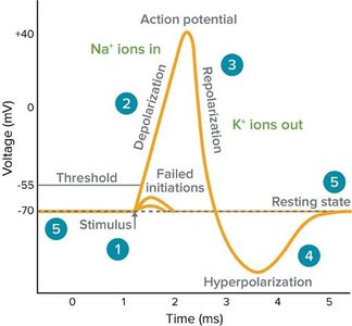
An action potential is a brief reversal of membrane potential (~100 mV) that occurs in excitable cells (neurons and muscle cells). It is the principal mechanism for long-distance neural communication and does not decay in amplitude with distance.
Occurs only in axons of neurons.
Also referred to as a nerve impulse.
Generated by voltage-gated ion channels responding to local currents (graded potentials).
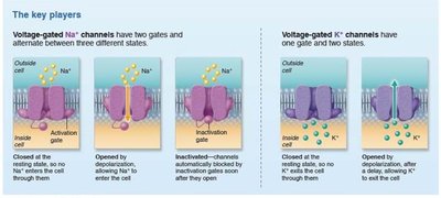
Phases of the Action Potential
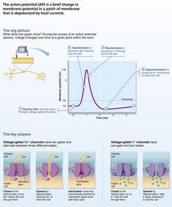
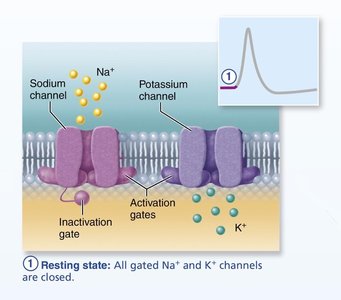
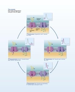
Resting State: All gated Na+ and K+ channels are closed; some K+ channels are open. Membrane potential is typically around -65 mV.
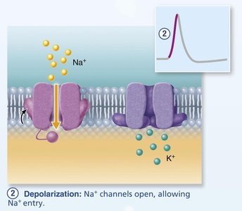
Depolarization: Na+ channels open, allowing Na+ influx; membrane potential becomes less negative and crosses threshold.
Rising Phase: Rapid opening of all Na+ channels; membrane potential spikes toward +30 mV.
Overshoot: Na+ channels inactivate; K+ channels open slowly.
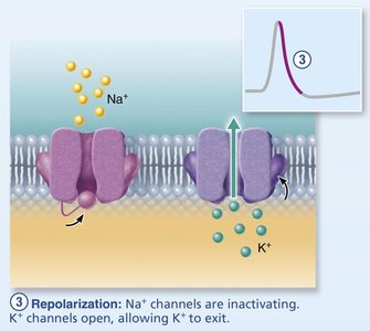
Repolarization: Na+ channels remain inactivated; K+ channels open, allowing K+ efflux; membrane potential returns toward resting.
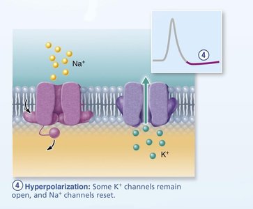
Hyperpolarization: Some K+ channels remain open; Na+ channels reset; membrane potential becomes more negative than resting.
Refractory Periods
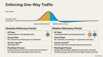
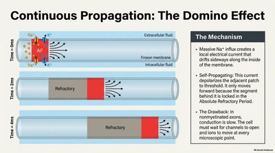
Absolute Refractory Period: During depolarization and early repolarization, Na+ channels are already open or inactivated; no new action potential can be initiated.
Relative Refractory Period: During late repolarization and hyperpolarization, K+ channels are still open; a stronger stimulus can initiate another action potential.
Propagation of Action Potentials
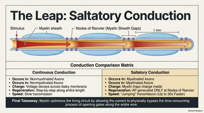
Continuous Propagation: Occurs in unmyelinated axons; step-by-step depolarization along the entire length; slow transmission.
Saltatory Conduction: Occurs in myelinated axons; action potential jumps from node to node (Nodes of Ranvier); much faster transmission (up to 30x faster).
The Synapse
Chemical Synapses
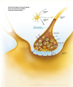
Synapses are specialized junctions where neurons communicate with other neurons or effector cells. The most common type is the chemical synapse, which uses neurotransmitters to transmit signals across a fluid-filled synaptic cleft.
Presynaptic neuron: Contains synaptic vesicles filled with neurotransmitter.
Postsynaptic neuron: Has receptor regions (usually on dendrites or cell body) that receive neurotransmitter.
Postsynaptic Potentials
Neurotransmitter binding to postsynaptic receptors generates graded potentials:
EPSP (Excitatory Postsynaptic Potential): Depolarizes the postsynaptic membrane, increasing the likelihood of action potential generation.
IPSP (Inhibitory Postsynaptic Potential): Hyperpolarizes the postsynaptic membrane, decreasing the likelihood of action potential generation.
Neurotransmitter Receptors
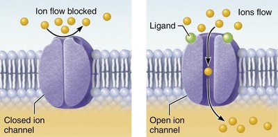
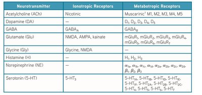
Ionotropic receptors: Ligand-gated ion channels; mediate rapid synaptic transmission; selective for specific ions.
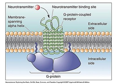
Metabotropic receptors: G-protein-coupled receptors; responses are indirect, complex, slow, and prolonged; activate second messenger systems.
Neurotransmitters
Neurotransmitters are the chemical messengers of the nervous system. Over 50 have been identified and are classified chemically and functionally. Key examples include:
Glutamate: Generally excitatory.
GABA: Generally inhibitory.
Acetylcholine (ACh): Excitatory and inhibitory.
Norepinephrine: Excitatory and inhibitory.
Excitatory Synapses and EPSPs
Neurotransmitter binding opens chemically gated channels, allowing Na+ influx and K+ efflux. The net effect is depolarization (EPSP). If the EPSP is strong enough to reach the axon hillock, an action potential is generated.
Inhibitory Synapses and IPSPs
Neurotransmitter binding opens channels for K+ or Cl–, causing hyperpolarization (IPSP). This moves the membrane potential farther from threshold, reducing the likelihood of action potential generation.
Summation of Postsynaptic Potentials
Integration and Modification
A single EPSP cannot induce an action potential, but multiple EPSPs can summate to reach threshold. IPSPs can also summate, and most neurons receive both excitatory and inhibitory inputs.
Temporal summation: Rapid-fire impulses from one or more presynaptic neurons add together.
Spatial summation: Simultaneous stimulation by multiple terminals; many receptors activated, EPSPs add together.
Comparison of Synaptic Potentials and Action Potentials
Property | Synaptic Potential | Action Potential |
|---|---|---|
Amplitude | Variable, graded | All-or-none, fixed |
Decay | Decays with distance | Does not decay |
Location | Dendrites/cell body | Axon |
Initiation | Neurotransmitter binding | Threshold depolarization |
