Indietro
Nerve Signaling and Membrane Potentials in the Nervous System
Guida di studio - Note intelligenti
 Appunti personalizzati basati sui tuoi materiali, ampliati con definizioni chiave, esempi e contesto.
Nerve Signaling and Membrane Potentials
Resting Membrane Potential (RMP)
The resting membrane potential (RMP) is the electrical charge difference across the plasma membrane of a neuron when it is not actively sending a signal. This potential is essential for the generation and propagation of nerve impulses.
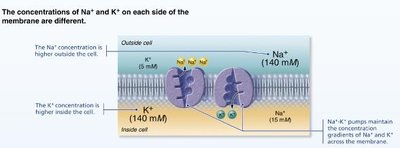
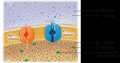
Ion Distribution: The RMP is primarily established by the unequal distribution of sodium (Na+) and potassium (K+) ions across the cell membrane.
Na+: Concentration is much higher outside the cell.
K+: Concentration is much higher inside the cell.
Negative Anions: Large negatively charged molecules (e.g., proteins, phosphates) are more abundant inside the cell, contributing to the negative charge.
Polarization: The cell membrane is said to be polarized due to this charge difference.
Maintaining Resting Membrane Potential
Leakage of Na+ and K+ ions through their respective channels tends to disrupt the RMP. The sodium-potassium pump (Na+/K+ ATPase) is crucial for maintaining this potential.
Na+/K+ Pump: Actively transports 3 Na+ ions out of the cell and 2 K+ ions into the cell, using ATP.
Result: Maintains high Na+ outside and high K+ inside, preserving the negative internal environment.
Nerve Signaling: Ion Channels
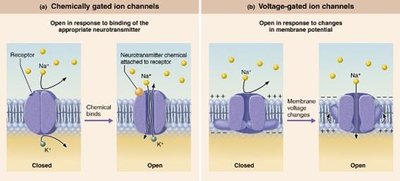
Nerve signaling depends on the movement of ions through specialized channels in the plasma membrane. There are two main types:
Ligand-gated (chemically gated) channels: Open in response to binding of a specific chemical (e.g., neurotransmitter).
Voltage-gated channels: Open in response to changes in membrane potential.
Action Potential: Generation and Phases
1. Resting State
All voltage-gated Na+ and K+ channels are closed. Only leakage channels are open, maintaining the RMP.
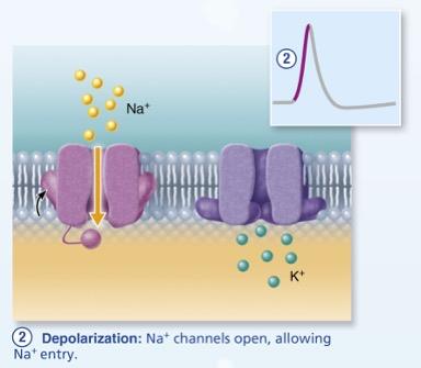
2. Depolarization
When a stimulus reaches the neuron, voltage-gated Na+ channels open, allowing Na+ to rush into the cell. This causes the inside of the membrane to become less negative (depolarization).
Threshold: If the membrane potential reaches about -55 to -50 mV, all Na+ channels open in a positive feedback loop.
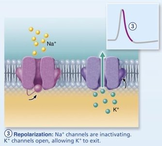
3. Repolarization
Na+ channels inactivate and voltage-gated K+ channels open. K+ exits the cell, restoring the negative internal charge (repolarization).
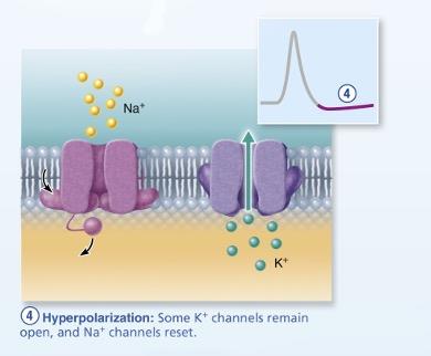
4. Hyperpolarization
Some K+ channels remain open, causing the membrane potential to become even more negative than the resting state. Na+ channels reset during this phase.
Summary Table: Phases of the Action Potential
Phase | Main Ion Movement | Membrane Potential Change |
|---|---|---|
Resting | Leakage of K+ and Na+ | -70 mV (stable) |
Depolarization | Na+ influx | Becomes less negative |
Repolarization | K+ efflux | Returns to negative |
Hyperpolarization | Continued K+ efflux | More negative than resting |
Conducting a Nerve Signal
Propagation of Action Potentials
An action potential at one region of the axon triggers a wave of depolarization along the axon, known as a nerve signal. After an action potential, the membrane enters a refractory period during which it cannot be re-excited immediately.
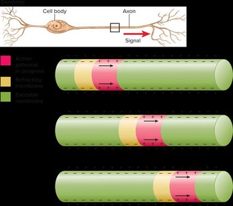
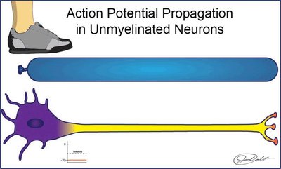
Unmyelinated Neurons: Continuous Conduction
In unmyelinated axons, voltage-gated Na+ and K+ channels are distributed along the entire length of the axon. The action potential is regenerated at every point along the axon, resulting in continuous conduction.
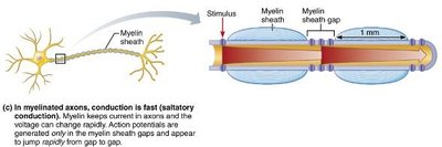
Myelinated Neurons: Saltatory Conduction
Myelinated axons have few voltage-gated channels in the myelinated regions (internodes), but many at the nodes of Ranvier. The action potential appears to jump from node to node, a process called saltatory conduction, which is much faster and more energy-efficient than continuous conduction.
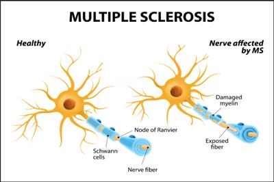
Clinical Homeostatic Imbalance: Multiple Sclerosis (MS)
Multiple sclerosis (MS) is an autoimmune disease in which the immune system attacks the myelin sheaths of neurons in the central nervous system. This disrupts saltatory conduction, leading to a variety of neurological symptoms.
Symptoms: Visual disturbances, muscle weakness, loss of coordination, speech difficulties, incontinence.
Treatment: Immune-modifying drugs; high vitamin D levels may reduce risk.
Transmitting a Nerve Signal to the Next Cell
Types of Synapses
Electrical synapses: Direct cytoplasmic connections via gap junctions; rapid communication; rare in adults.
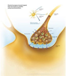
Chemical synapses: Most common; involve neurotransmitter release across a synaptic cleft.
Chemical Synapse Structure and Function
When an action potential reaches the axon terminal of the presynaptic neuron, it triggers the release of neurotransmitters from synaptic vesicles into the synaptic cleft. These neurotransmitters bind to receptors on the postsynaptic neuron, initiating a new electrical signal.
Fate of Neurotransmitters
Neurotransmitters are either broken down by enzymes or reabsorbed by the presynaptic neuron.
Examples include acetylcholine (ACh), norepinephrine, serotonin, and dopamine.
Clinical Homeostatic Imbalance: Parkinson’s Disease
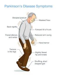
Parkinson’s disease is a progressive neurodegenerative disorder characterized by the loss of dopamine-releasing neurons in the brain. Dopamine normally acts as an inhibitory neurotransmitter, preventing excessive motor activity. Its loss leads to involuntary muscle contractions and motor symptoms.
Symptoms: Tremors, rigidity, bradykinesia (slowness of movement), postural instability.