 Indietro
Nervous System and Special Senses Study Guide – Anatomy & Physiology
Guida di studio - Note intelligenti
 Appunti personalizzati basati sui tuoi materiali, ampliati con definizioni chiave, esempi e contesto.
Q287. Using Figure 8.1, identify the auricle (pinna).
Background
Topic: Anatomy of the Ear
This question tests your ability to recognize the external structures of the ear, specifically the auricle (pinna), which is the visible part of the ear that collects sound waves.
Key Terms:
Auricle (pinna): The external, cartilaginous part of the ear.
External ear: Includes the auricle and external acoustic meatus.
Step-by-Step Guidance
Examine Figure 8.1 and locate the structure that forms the outermost visible part of the ear.
Identify which label points to the curved, cartilaginous area that is responsible for funneling sound into the ear canal.
Compare the labels to the anatomical position of the auricle (pinna).
Try solving on your own before revealing the answer!
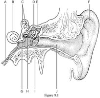
Final Answer: Label F
The auricle (pinna) is indicated by Label F in Figure 8.1. This is the external, visible part of the ear that collects sound waves.
Q288. Using Figure 8.1, identify the tympanic membrane.
Background
Topic: Anatomy of the Ear
This question tests your ability to locate the tympanic membrane (eardrum), which separates the external ear from the middle ear and vibrates in response to sound waves.
Key Terms:
Tympanic membrane: Also known as the eardrum, a thin membrane that transmits sound vibrations.
Middle ear: The area just beyond the tympanic membrane.
Step-by-Step Guidance
Look at Figure 8.1 and find the structure that forms a boundary between the external auditory canal and the middle ear cavity.
Identify which label points to the thin, oval-shaped membrane.
Compare the labels to the anatomical position of the tympanic membrane.
Try solving on your own before revealing the answer!
Final Answer: Label D
The tympanic membrane is indicated by Label D in Figure 8.1. It is the membrane that vibrates in response to sound waves and separates the external ear from the middle ear.
Q376. Using Figure 7.1, identify the neural processes that convey incoming messages toward the cell body.
Background
Topic: Neuron Structure
This question tests your understanding of neuron anatomy, specifically the dendrites, which receive signals and transmit them toward the cell body.
Key Terms:
Dendrites: Branch-like extensions that receive incoming signals.
Cell body (soma): The main part of the neuron containing the nucleus.
Step-by-Step Guidance
Examine Figure 7.1 and locate the structures that branch out from the cell body.
Identify which label points to the extensions that are responsible for receiving incoming messages.
Compare the labels to the anatomical position of the dendrites.
Try solving on your own before revealing the answer!
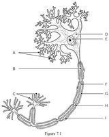
Final Answer: Label H
The neural processes that convey incoming messages toward the cell body (dendrites) are indicated by Label H in Figure 7.1.
Q377. Using Figure 7.1, identify the metabolic center of the neuron.
Background
Topic: Neuron Structure
This question tests your ability to identify the cell body (soma) of a neuron, which contains the nucleus and is responsible for metabolic activities.
Key Terms:
Cell body (soma): The central part of the neuron containing the nucleus.
Nucleus: The organelle that controls cell metabolism and function.
Step-by-Step Guidance
Examine Figure 7.1 and locate the large, central area of the neuron.
Identify which label points to the region containing the nucleus.
Compare the labels to the anatomical position of the cell body.
Try solving on your own before revealing the answer!
Final Answer: Label D
The metabolic center of the neuron (cell body/soma) is indicated by Label D in Figure 7.1.
