 Indietro
Nervous System, Central Nervous System, and Special Senses: Study Guide
Guida di studio - Note intelligenti
 Appunti personalizzati basati sui tuoi materiali, ampliati con definizioni chiave, esempi e contesto.
Nervous System (Ch. 11)
Neural Tissue: Glial Cells and Neurons
The nervous system is composed of specialized cells called neurons and supporting cells known as glial cells. Glial cells provide structural and functional support to neurons, and are classified based on their location in the central nervous system (CNS) or peripheral nervous system (PNS).
Glial Cells in the CNS:
Astrocytes: Maintain the blood-brain barrier, regulate ion concentration, and provide structural support.
Microglia: Act as immune cells, removing debris and pathogens via phagocytosis.
Oligodendrocytes: Form myelin sheaths around CNS axons, increasing conduction speed.
Ependymal Cells: Line ventricles and produce cerebrospinal fluid (CSF).
Glial Cells in the PNS:
Schwann Cells: Form myelin sheaths around PNS axons, aid in axon repair.
Saltatory Conduction: Rapid transmission of action potentials along myelinated axons, jumping from node to node.
Axon/Nerve Repair: Schwann cells facilitate regeneration of damaged axons in the PNS.
Neurons: General Anatomy & Classification
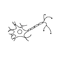
Neurons are the functional units of the nervous system, specialized for transmitting electrical and chemical signals. They are classified by structure and function.
Structural Types: Multipolar, bipolar, and unipolar neurons.
Functional Types: Sensory (afferent), motor (efferent), and interneurons.
Synapses: Structure and Function
A synapse is the junction between two neurons, where information is transmitted from the presynaptic neuron to the postsynaptic neuron via neurotransmitters.
Presynaptic Membrane: Releases neurotransmitters into the synaptic cleft.
Postsynaptic Membrane: Contains receptors for neurotransmitters, initiating a response.
Fates of Neurotransmitter: Can be degraded, reabsorbed, or bind to receptors to elicit effects.
Membrane Potentials
Neurons maintain a resting membrane potential due to differences in ion concentrations across the membrane. This potential is essential for the generation of electrical signals.
Resting Membrane Potential: Typically -70 mV, maintained by Na+/K+ pumps and ion channels.
Key Ions: Sodium (Na+), Potassium (K+), Chloride (Cl-).
Graded Potentials vs. Action Potentials
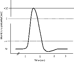
Graded potentials are local changes in membrane potential, while action potentials are all-or-none electrical impulses that propagate along axons.
Graded Potentials: Vary in magnitude, decay with distance, occur in dendrites and cell bodies.
Action Potentials: Fixed magnitude, propagate without decrement, occur in axons.
Neural Summation
Neural summation refers to the process by which multiple synaptic inputs combine to influence the postsynaptic neuron.
Temporal Summation: Multiple signals from one neuron in rapid succession.
Spatial Summation: Signals from multiple neurons at the same time.
Action Potentials: Refractory Periods and Frequency
Action potentials have absolute and relative refractory periods, which affect the frequency of nerve impulses.
Absolute Refractory Period: No new action potential can be generated.
Relative Refractory Period: A stronger stimulus can initiate another action potential.
Central Nervous System (Ch. 12)
CNS/PNS Organization
The nervous system is divided into the central nervous system (CNS) and peripheral nervous system (PNS). The CNS includes the brain and spinal cord, while the PNS consists of nerves and ganglia outside the CNS.
Anatomy of the Brain: Lobes and Functions
The brain is divided into several lobes, each with distinct functions:
Frontal Lobe: Motor control, problem-solving, speech production.
Parietal Lobe: Sensory processing, spatial orientation.
Temporal Lobe: Auditory processing, memory, language comprehension.
Occipital Lobe: Visual processing.
Insular Lobe: Taste, visceral sensation, emotion.
White Matter Tracts
Commissural Tracts: Connect corresponding areas of the two hemispheres (e.g., corpus callosum).
Association Tracts: Connect different regions within the same hemisphere.
Projection Tracts: Connect the cortex with lower brain regions and spinal cord.
Nuclei: Cortical and Basal Nuclei
Cortical Nuclei: Groups of neurons in the cortex responsible for primary sensory and motor functions.
Basal Nuclei: Involved in motor control and coordination.
Diencephalon: Parts and Functions
Epithalamus: Contains pineal gland, regulates sleep-wake cycles.
Thalamus: Relay station for sensory information.
Hypothalamus: Controls autonomic functions, hormone release, homeostasis.
Brain Stem: Parts and Functions
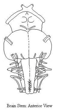
The brain stem consists of the midbrain, pons, and medulla oblongata, and is responsible for basic life functions such as breathing, heart rate, and reflexes.
Cerebellum: Motor Programs and Control
The cerebellum coordinates voluntary movements, balance, and motor learning.
Cerebrospinal Fluid (CSF)
CSF is produced by the choroid plexus in the ventricles, circulates through the CNS, and is drained via arachnoid villi. It cushions the brain and removes waste.
Meninges
The meninges are three protective layers surrounding the brain and spinal cord: dura mater, arachnoid mater, and pia mater.
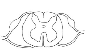
Anatomy of the Spinal Cord: Sensory and Motor Pathways
The spinal cord transmits sensory and motor information between the body and brain. Sensory pathways are located dorsally, motor pathways ventrally.
Reflex Arc
A reflex arc is the neural pathway that mediates a reflex action, typically involving a sensory neuron, interneuron, and motor neuron.
Special Senses (Ch. 15)
Vision: Anatomy and Physiology
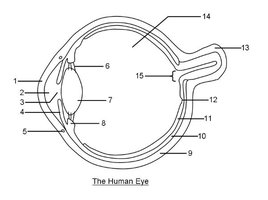
The eye is composed of accessory structures and three main tunics (layers): fibrous, vascular, and neural. Each layer has distinct functions in vision.
Accessory Structures: Eyelids, eyelashes, lacrimal apparatus, protect and lubricate the eye.
Tunics/Layers:
Fibrous Tunic: Sclera and cornea, provide shape and protection.
Vascular Tunic: Choroid, ciliary body, iris; supply blood and control light entry.
Neural Tunic: Retina; contains photoreceptors (rods and cones).
Iris and Lens: Control light entry and focus.
Aqueous Humor: Maintains intraocular pressure, nourishes lens and cornea.
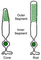
Visual Perception: Rods and Cones
Rods and cones are photoreceptors in the retina responsible for vision. Rods detect dim light and provide black-and-white vision, while cones detect color and function in bright light.
Neural Pathway and Physiology of Vision
Visual information is transmitted from the retina to the brain via the optic nerve, processed in the visual cortex.
Autonomic Regulation: Controls iris and pupil size.
Lens and Pupil Accommodation: Adjusts focus for near and far objects.
Hearing: Anatomy and Physiology
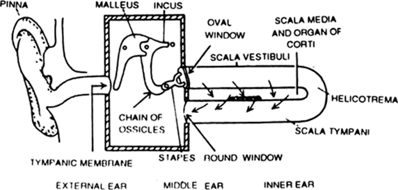
The ear is divided into external, middle, and inner regions, each with specialized structures for hearing and balance.
External Ear: Pinna, external auditory canal, tympanic membrane; collects sound.
Middle Ear: Ossicles (malleus, incus, stapes), pharyngotympanic tube; transmit and amplify sound.
Inner Ear: Cochlea (hearing), vestibule and semicircular canals (equilibrium).
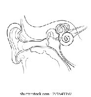
Inner Ear: Labyrinth and Equilibrium
The inner ear contains the labyrinth, which is responsible for balance and spatial orientation.
Vestibule: Detects static equilibrium.
Saccule and Utricle: Detect linear acceleration.
Otoliths: Calcium carbonate crystals involved in sensing gravity.
Semicircular Canals: Detect rotational movement (dynamic equilibrium).
Cochlea: Sound Detection and Coding
The cochlea is a spiral-shaped organ that converts sound waves into electrical signals. It contains the organ of Corti, hair cells, and fluid-filled chambers (perilymph and endolymph).
Amplitude: Determines loudness.
Pitch: Determined by frequency of sound waves.
Hair Cells: Specialized for bimodal signaling, convert mechanical vibrations to nerve impulses.
Sensory Neural Pathway: Auditory nerve transmits signals to the auditory cortex.
Additional info: The notes have been expanded with academic context to ensure completeness and clarity for exam preparation.
