 Indietro
E2 Lecture 4: Nervous System Pt. 2
Guida di studio - Note intelligenti
 Appunti personalizzati basati sui tuoi materiali, ampliati con definizioni chiave, esempi e contesto.
Nervous System: Electrical Properties and Signaling
Resting Membrane Potential (RMP)
The resting membrane potential (RMP) is the voltage difference across the cell membrane of a neuron when it is not actively transmitting a signal. This potential is essential for the function of excitable cells such as neurons and muscle cells.
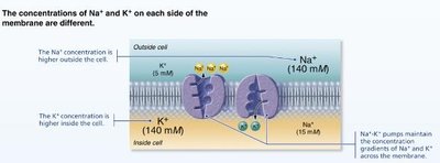
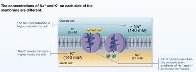
Ion Distribution: The RMP is established by the unequal distribution of ions, mainly sodium (Na+) and potassium (K+), across the cell membrane.
Outside the Cell: Na+ is much more concentrated outside the cell membrane.
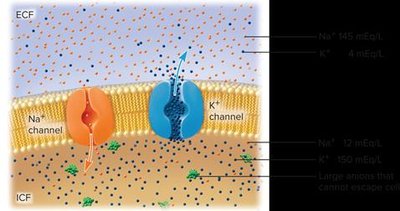
Inside the Cell: K+ is much more concentrated inside the cell, along with negatively charged anions (phosphates, proteins, nucleic acids).
Polarization: The interior of the cell is more negatively charged than the exterior, resulting in a polarized membrane.
Maintaining Resting Membrane Potential
The RMP is maintained by selective permeability of the membrane and the activity of the sodium-potassium pump (Na+/K+ ATPase).
Leak Channels: Na+ and K+ channels allow ions to leak across the membrane, which could disrupt the RMP.
Sodium-Potassium Pump: This pump actively transports 3 Na+ ions out of the cell and 2 K+ ions into the cell, using ATP. This maintains the concentration gradients and the negative interior.
Equation: The pump's activity can be summarized as:
Nerve Signaling: Ion Channels
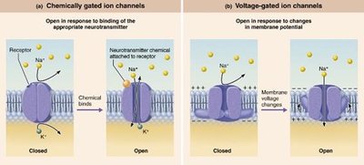
Nerve signaling depends on the opening and closing of ion channels in the plasma membrane. These channels are crucial for generating and propagating electrical signals.
Ligand-Gated Channels: Open in response to binding of a specific chemical (neurotransmitter).
Voltage-Gated Channels: Open in response to changes in membrane potential.
Action Potential: Generation and Phases
Step 1: Resting State
At rest, all voltage-gated Na+ and K+ channels are closed. Only leakage channels are open, maintaining the RMP.
Na+ Channels: Have two gates (activation and inactivation).
K+ Channels: Have one voltage-sensitive gate.
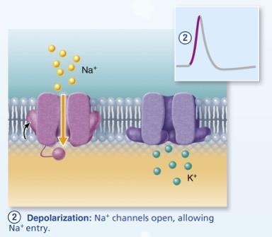
Step 2: Depolarization
Depolarization occurs when voltage-gated Na+ channels open, allowing Na+ to rush into the cell. This causes the membrane potential to become less negative.
Threshold: When the membrane potential reaches -55 to -50 mV, all Na+ channels open due to positive feedback.
Result: Rapid influx of Na+ leads to a spike in membrane potential.
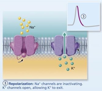
Step 3: Repolarization
Repolarization restores the negative membrane potential. Na+ channels inactivate, and voltage-gated K+ channels open, allowing K+ to exit the cell.
Result: The membrane potential returns toward the resting state.
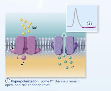
Step 4: Hyperpolarization
Some K+ channels remain open, causing excessive K+ efflux. The inside of the membrane becomes more negative than the resting state, resulting in hyperpolarization.
Na+ Channels: Begin to reset during this phase.
Conducting a Nerve Signal
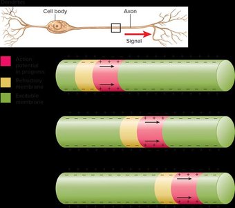
Propagation of Action Potentials
An action potential in one area of the membrane triggers a domino effect, causing action potentials to propagate down the axon. This is the basis of nerve signaling.
Refractory Period: After an action potential, the affected area cannot be stimulated again for a short time.
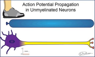
Unmyelinated Neurons: Continuous Conduction
In unmyelinated axons, voltage-gated Na+ and K+ channels are distributed along the entire axon. Action potentials occur at every point, resulting in continuous conduction.
Speed: Slower than myelinated conduction.
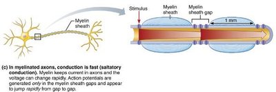
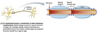
Myelinated Neurons: Saltatory Conduction
Myelinated axons have few voltage-gated channels in the myelin-covered internodes. Action potentials are generated only at the nodes of Ranvier, where ion channels are concentrated. The signal appears to jump from node to node, a process called saltatory conduction.
Speed: Much faster than continuous conduction.
Energy Efficiency: Less energy is required due to fewer ion exchanges.
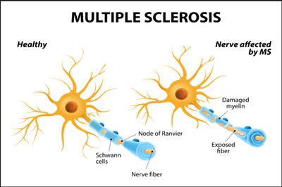
Clinical Homeostatic Imbalance: Multiple Sclerosis (MS)
Multiple sclerosis (MS) is an autoimmune disease that destroys myelin sheaths in the CNS, impairing nerve conduction.
Symptoms: Visual disturbances, weakness, loss of muscular control, speech disturbances, incontinence.
Treatment: Drugs that modify immune system activity; maintaining high blood levels of vitamin D may reduce risk.
Transmitting a Nerve Signal to the Next Cell
Types of Synapses
Nerve signals are transmitted to the next cell via synapses, which can be electrical or chemical.
Electrical Synapses: Less common; joined by gap junctions for rapid communication, most abundant in embryonic nervous tissue.
Chemical Synapses: Most common; involve neurotransmitter release.
Chemical Synaptic Transmission
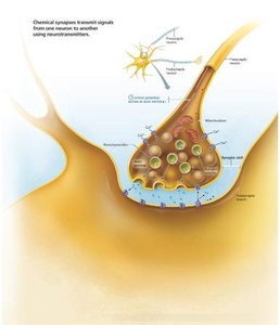
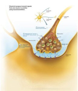
When a wave of action potentials reaches the axon terminal, neurotransmitters are released into the synaptic cleft.
Presynaptic Neuron: The cell sending the signal.
Postsynaptic Neuron: The cell receiving the signal.
Synaptic Cleft: The gap between the two neurons.
Synaptic Vesicles: Contain neurotransmitters; undergo exocytosis upon arrival of nerve signal.
Fate of Neurotransmitters
After release, neurotransmitters are either broken down by enzymes or reabsorbed by the presynaptic neuron.
Examples: Acetylcholine (ACh), norepinephrine, serotonin, dopamine.
Number: More than 100 known neurotransmitters.
Clinical Homeostatic Imbalance: Parkinson’s Disease
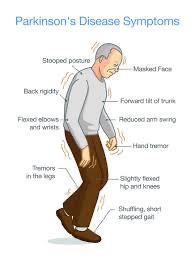
Parkinson’s disease is characterized by progressive loss of motor control, typically beginning in a person's 50s or 60s. It is caused by degeneration of dopamine-releasing neurons.
Dopamine: Acts as an inhibitory neurotransmitter, preventing excessive activity. Loss leads to involuntary muscle contractions.
Symptoms: Tremors, rigidity, shuffling gait, stooped posture, masked face.
Summary Table: Ion Distribution and Membrane Potential
Location | Na+ Concentration | K+ Concentration | Charge |
|---|---|---|---|
Outside Cell | High (140 mM) | Low (5 mM) | Positive |
Inside Cell | Low (15 mM) | High (140 mM) | Negative |
Additional info: The sodium-potassium pump and selective permeability are fundamental for maintaining the RMP and enabling rapid signaling in neurons.
