Indietro
Nervous System: Organization, Structure, and Function
Guida di studio - Note intelligenti
 Appunti personalizzati basati sui tuoi materiali, ampliati con definizioni chiave, esempi e contesto.
Nervous System: Organization and Structure
Introduction to the Nervous System
The nervous system is the primary communication and control system of the body, responsible for detecting changes, processing information, and coordinating rapid responses. It works closely with the endocrine system to maintain homeostasis.
Communication: Detects internal and external changes (stimuli).
Integration: Processes and interprets sensory input.
Response: Activates muscles and glands for motor output.
Anatomical Divisions of the Nervous System
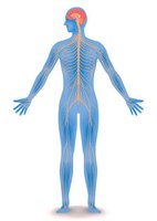
The nervous system is divided into two major anatomical divisions:
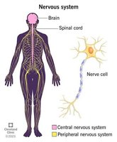
Central Nervous System (CNS): Consists of the brain and spinal cord; primary center for processing and integrating information.
Peripheral Nervous System (PNS): Composed of nerves and ganglia outside the CNS; carries sensory information to the CNS and motor commands away from it.
Functional Divisions of the Peripheral Nervous System
The PNS is functionally divided into sensory and motor divisions:
Sensory (Afferent) Division: Carries information from sensory receptors toward the CNS.
Motor (Efferent) Division: Carries commands from the CNS to muscles and glands. Subdivided into:
Somatic Nervous System: Controls voluntary movements of skeletal muscles.
Autonomic Nervous System: Regulates involuntary activities of cardiac muscle, smooth muscle, and glands.
Autonomic Nervous System: Sympathetic vs Parasympathetic
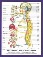
The autonomic nervous system is divided into:
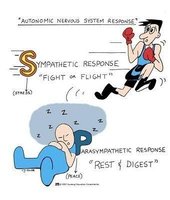
Sympathetic Division: Prepares the body for 'fight or flight' responses; increases heart rate, dilates airways, and reduces digestive activity.
Parasympathetic Division: Promotes 'rest and digest' functions; slows heart rate, stimulates digestion, and conserves energy.
Nervous Tissue
Cell Types in Nervous Tissue
Nervous tissue consists of two major categories of cells:
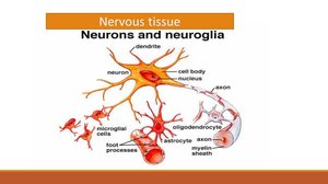
Neurons: Excitable cells that receive, process, and transmit information using electrical and chemical signals.
Neuroglia: Support, protect, and nourish neurons; maintain the environment for neural activity.
Neuroglia of the Central Nervous System
Four major types of neuroglia in the CNS:
Astrocytes: Provide structural support and maintain the blood-brain barrier.
Oligodendrocytes: Produce myelin sheaths around CNS axons.
Microglia: Act as immune defense, removing pathogens and debris.
Ependymal Cells: Line ventricles and central canal; produce and circulate cerebrospinal fluid (CSF).
Neuroglia of the Peripheral Nervous System
Two major types of neuroglia in the PNS:
Schwann Cells: Form myelin sheaths around peripheral axons.
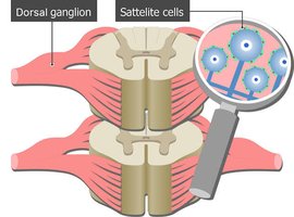
Satellite Cells: Surround neuron cell bodies in ganglia, providing support and regulating chemical environment.
Myelination: Oligodendrocytes vs Schwann Cells
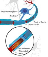
Oligodendrocytes: Myelinate axons in the CNS by wrapping their processes around axons.
Schwann Cells: Myelinate axons in the PNS by wrapping their own cell membrane repeatedly around the axon.
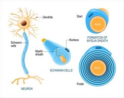
Structure and Classification of Neurons
Structure of a Neuron
Neurons are specialized for receiving and transmitting information.
Dendrites: Receive signals from other neurons or sensory receptors.
Cell Body (Soma): Contains nucleus and organelles; metabolic center.
Axon Hillock: Region where action potentials are initiated.
Axon: Conducts action potentials away from the cell body.
Axon Terminal: Releases neurotransmitters to communicate with other cells.
Myelin Sheath: Insulates axon, increases speed of impulse conduction.
Nodes of Ranvier: Gaps in myelin sheath; facilitate saltatory conduction.
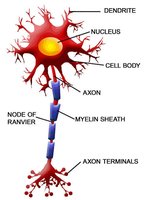
Structural Classification of Neurons
Neurons are classified by their structure:
Multipolar Neurons: One axon, multiple dendrites; most common type.
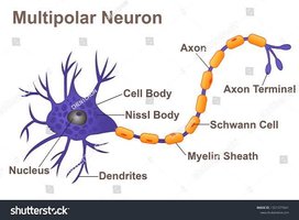
Bipolar Neurons: One axon, one dendrite; found in special sensory pathways.
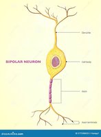
Unipolar Neurons: One process from cell body; rare in humans, common in invertebrates.
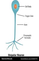
Pseudounipolar Neurons: One process splits into two branches; common in dorsal root ganglia.
Functional Classification of Neurons
Neurons are also classified by function:
Sensory (Afferent) Neurons: Carry information from receptors to CNS.
Motor (Efferent) Neurons: Carry impulses from CNS to muscles or glands.
Interneurons: Located in CNS; connect sensory and motor neurons, important for integration and reflexes.
Electrical Properties of Neurons
Resting Membrane Potential
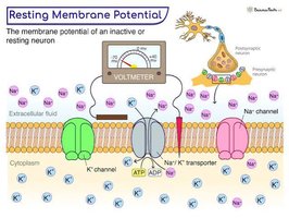
The resting membrane potential is the electrical difference across the neuron's membrane when not transmitting an impulse.
Typical Value: Approximately -70 mV (inside negative relative to outside).
Cause: Unequal distribution of ions and selective permeability of the membrane.
Ion Distribution and the Sodium-Potassium Pump
Sodium (Na+): Higher concentration outside the neuron.
Potassium (K+): Higher concentration inside the neuron.
Leak Channels: Allow passive movement of ions.
Sodium-Potassium Pump: Maintains gradients by pumping 3 Na+ out and 2 K+ in per ATP used.
Types of Ion Channels
Leak Channels: Always open; allow passive ion movement.
Ligand-Gated Channels: Open in response to neurotransmitter binding.
Voltage-Gated Channels: Open/close in response to changes in membrane potential.
Graded Potentials
Graded potentials are small, localized changes in membrane potential.
Depolarizing: Inside becomes less negative (Na+ influx).
Hyperpolarizing: Inside becomes more negative (K+ efflux or Cl- influx).
Summation of Graded Potentials
Temporal Summation: Rapid, repeated signals from one neuron.
Spatial Summation: Multiple neurons stimulate different locations simultaneously.
Threshold and Action Potential Initiation
Threshold: Critical membrane potential (~ -55 mV) required to initiate an action potential.
Action Potential: Rapid electrical change propagating along the axon.
Phases of an Action Potential
Depolarization: Voltage-gated Na+ channels open; Na+ influx; membrane potential rises to +30 mV.

Repolarization: Na+ channels inactivate; K+ channels open; K+ efflux restores negativity.
After-Hyperpolarization: Membrane becomes more negative than resting potential.
All-or-None Principle
Once threshold is reached, a full action potential occurs.
Strength of stimulus affects frequency, not amplitude.
Refractory Periods
Absolute Refractory Period: No new action potential can occur.
Relative Refractory Period: New action potential possible with stronger stimulus.
Conduction of Action Potentials
Continuous Conduction: Occurs in unmyelinated axons; slower.
Saltatory Conduction: Occurs in myelinated axons; action potential jumps between nodes of Ranvier; faster and more efficient.
Synaptic Communication
Types of Synapses
Electrical Synapse: Direct ion passage via gap junctions.
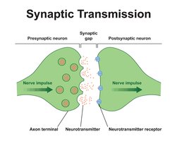
Chemical Synapse: Neurotransmitter release across synaptic cleft; most common in humans.
Structure of a Chemical Synapse
Presynaptic Axon Terminal: Contains synaptic vesicles and voltage-gated calcium channels.
Synaptic Cleft: Small gap between cells.
Postsynaptic Membrane: Contains receptors for neurotransmitters.
Steps of Chemical Synaptic Transmission
Action potential arrives at presynaptic terminal.
Depolarization opens voltage-gated Ca2+ channels.
Ca2+ triggers neurotransmitter release by exocytosis.
Neurotransmitter diffuses across synaptic cleft and binds to postsynaptic receptors.
Postsynaptic Potentials
Excitatory Postsynaptic Potential (EPSP): Depolarizes membrane, increases likelihood of action potential.
Inhibitory Postsynaptic Potential (IPSP): Hyperpolarizes membrane, decreases likelihood of action potential.
Summation and Cancellation
EPSPs and IPSPs combine via summation; if excitatory input exceeds inhibitory, threshold may be reached.
EPSP-IPSP cancellation occurs when excitatory and inhibitory inputs are equal.
Neurotransmitters and Receptors
Types of Neurotransmitter Receptors
Ionotropic Receptors: Direct action; receptor is part of ligand-gated ion channel.
Metabotropic Receptors: Indirect action; receptor activates G protein and intracellular signaling pathways.
Termination of Neurotransmitter Activity
Reuptake: Transport proteins remove neurotransmitter from synaptic cleft.
Enzymatic Degradation: Enzymes break down neurotransmitter (e.g., acetylcholinesterase).
Diffusion: Neurotransmitter diffuses away from synaptic cleft.
Synaptic Plasticity and Neural Circuits
Synaptic Plasticity
Synaptic plasticity is the ability of synapses to strengthen or weaken in response to activity, crucial for learning and memory.
Long-Term Potentiation (LTP): Strengthening of synaptic communication.
Long-Term Depression (LDP): Weakening of synaptic communication.
Types of Neural Circuits
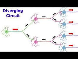
Diverging Circuit: One neuron communicates with multiple neurons, spreading information.
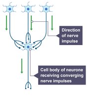
Converging Circuit: Multiple neurons communicate with one neuron, integrating information.
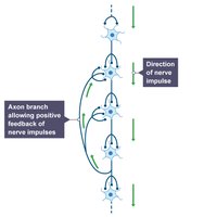
Reverberating Circuit: Feedback connections allow repeated stimulation and rhythmic responses.
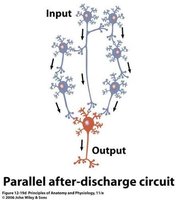
Parallel After-Discharge Circuit: Input neuron stimulates several parallel chains, which reconverge on a single output neuron; allows continued response after stimulus ends.