 Indietro
Nervous Tissue and Physiology: Electrophysiology, Signal Transmission, and Neurotransmitters
Guida di studio - Note intelligenti
 Appunti personalizzati basati sui tuoi materiali, ampliati con definizioni chiave, esempi e contesto.
Nervous Tissue and Electrophysiology
Principles of Electricity in Neurons
Neurons utilize electrical principles to transmit signals. The movement of ions across membranes generates electrical currents, which are governed by fundamental laws of physics.
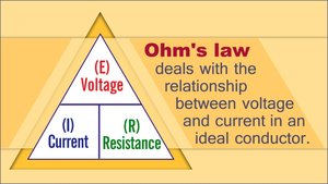
Voltage (V): The measure of potential energy created by separated electrical charges. It is always measured between two points; a larger difference means higher voltage. Units: volts (V), where 1000 mV = 1 V.
Current (I): The flow of electrical charge from one point to another.
Resistance (R): The hindrance to the flow of electrical charge. Insulators have high resistance, conductors have low resistance.
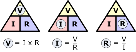
Ohm’s Law: Describes the relationship between voltage, current, and resistance: Current is proportional to voltage and inversely proportional to resistance.
Ion Channels and Membrane Potential
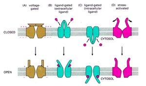
Ion channels are specialized proteins in the neuronal membrane that allow ions to move in and out, creating changes in membrane potential.
Chemically (Ligand) Gated Channels: Open when a specific chemical binds.
Voltage-Gated Channels: Open and close in response to changes in membrane potential.
Mechanically Gated Channels: Open by physical deformation of the membrane.
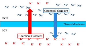
Ions diffuse down their concentration and electrical gradients, moving toward areas of opposite charge.
Resting Membrane Potential
The resting membrane potential is the difference in electrical charge across the neuronal membrane when the cell is at rest, typically around -70 mV.
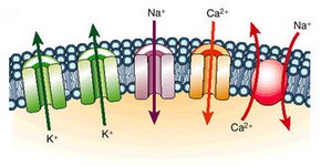
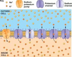
Na+/K+ Pump: Actively transports 3 Na+ out and 2 K+ in, maintaining the gradient.
Leak Channels: Allow passive diffusion of Na+ and K+, but there are more K+ leak channels, making the inside more negative.
Polarization: The outside is positively charged, the inside is negatively charged.
Ionic Composition: Cytosol has lower Na+ and higher K+; extracellular fluid has balanced cations and Cl-.
Changes in Membrane Potential
Neurons change their membrane potential in response to stimuli, which is essential for signal transmission.
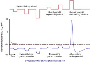
Depolarization: Membrane potential becomes less negative (e.g., -70 mV to -65 mV).
Hyperpolarization: Membrane potential becomes more negative (e.g., -70 mV to -75 mV).
Graded Potential: Local, short-distance changes in membrane potential.
Action Potential: Long-distance, all-or-nothing electrical signal.
Local and Action Potentials
Local Potentials
Local potentials are small, graded changes in membrane potential that occur in dendrites and the cell body. They are reversible and decremental, meaning they decrease in strength with distance.
Graded Depolarization: Sodium ions enter, making the cell less negative.
Graded Hyperpolarization: Chloride ions enter, making the cell more negative.
Caused by ligand-gated ion channels.
Useful for short-distance signaling.
Action Potentials
Action potentials are rapid, long-distance electrical signals generated in axons. They are all-or-nothing and do not decay with distance.
Generation:
Resting state: All gated Na+ and K+ channels closed.
Depolarization: Na+ channels open, threshold reached (~ -55 mV).
Repolarization: Na+ channels inactivate, K+ channels open.
Hyperpolarization: K+ channels remain open, Na+ channels reset.
Na+/K+ pumps restore resting potential.
Threshold: Action potential only occurs if threshold is reached.
Propagation: Spreads from point of origin; frequency determines stimulus intensity.
Refractory Periods
Refractory periods ensure unidirectional propagation and limit the frequency of action potentials.
Absolute Refractory Period: Neuron cannot respond; Na+ channels are inactivated.
Relative Refractory Period: Elevated threshold; K+ channels remain open.
Propagation of Action Potentials
Saltatory vs. Continuous Conduction
Action potentials propagate differently depending on whether the axon is myelinated or unmyelinated.
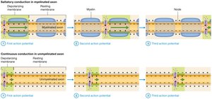
Saltatory Conduction: In myelinated axons, action potentials jump from node to node, increasing speed and efficiency.
Continuous Conduction: In unmyelinated axons, every section of the membrane must depolarize, slowing conduction.
Conduction Velocity
Axon Diameter: Larger axons conduct faster due to less resistance.
Degree of Myelination: More myelin increases conduction speed.
Fiber Groups:
Group A: Largest diameter, thick myelin, fastest (somatic sensory/motor fibers).
Group B: Intermediate diameter, light myelin, moderate speed (ANS).
Group C: Smallest diameter, non-myelinated, slowest (ANS).
Neuronal Synapses and Signal Transmission
Synapse Structure and Function
Synapses are junctions that mediate information transfer between neurons. The presynaptic neuron transmits the signal, while the postsynaptic neuron receives it.
Axodendritic Synapses: Between axon endings and dendrites.
Axosomatic Synapses: Between axon endings and cell body.
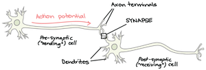
Electrical Synapses
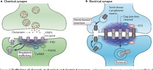
Electrical synapses are less common and consist of gap junctions, allowing direct flow of ions between cells. They are found in some brain regions and embryonic nervous tissue.
Chemical Synapses
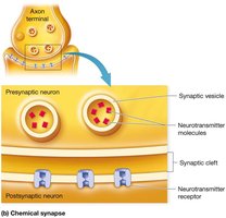
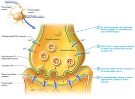
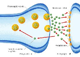
Chemical synapses are the most common type in the nervous system. They involve the release of neurotransmitters from synaptic vesicles in the presynaptic neuron into the synaptic cleft, where they bind to receptors on the postsynaptic neuron.
Synaptic Vesicles: Contain neurotransmitters.
Synaptic Cleft: Small gap separating neurons.
Neurotransmitter Receptors: Bind neurotransmitters to induce a response.
Events at Chemical Synapse
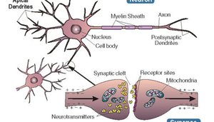
The process of signal transmission at a chemical synapse involves several steps:
Action potential arrives at presynaptic axon terminal.
Voltage-gated Ca2+ channels open, Ca2+ influx triggers neurotransmitter release.
Neurotransmitter diffuses across synaptic cleft and binds to postsynaptic receptors.
Ion channels open, leading to local potential and possibly action potential if threshold is reached.
Postsynaptic Potentials
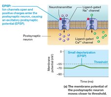
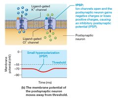
Postsynaptic potentials are local changes in membrane potential in the postsynaptic neuron.
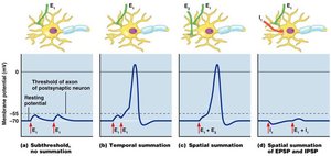
Excitatory Postsynaptic Potential (EPSP): Small local depolarization, moves membrane potential closer to threshold (Na+ or Ca2+ channels open).
Inhibitory Postsynaptic Potential (IPSP): Small local hyperpolarization, moves membrane potential farther from threshold (K+ or Cl- channels open).
Integration and Summation
Neurons integrate multiple signals through summation, which determines whether an action potential will be generated.
Temporal Summation: Rapid impulses from presynaptic neurons.
Spatial Summation: Large stimulation event, much neurotransmitter released.
Facilitated Neurons: Partially depolarized, more easily excited.
Synaptic Potentiation: Repeated use enhances synapse’s ability to be excited (long-term potentiation).
Presynaptic Inhibition: One neuron can inhibit the impulse of another.
Major Neurotransmitters
Types and Functions of Neurotransmitters
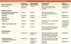
Neurotransmitters are chemical messengers that transmit signals across synapses. They can be excitatory or inhibitory, affecting the postsynaptic neuron in different ways.
Acetylcholine (ACh): Excitatory; found in neuromuscular junctions, brain, spinal cord, and autonomic nervous system.
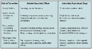
Norepinephrine: Influences heart rate, blood pressure, digestion; regulates sleep/wake cycle, attention, feeding behaviors.
Epinephrine: Similar to norepinephrine; more widely used as a hormone.
Dopamine: Movement coordination, emotion, motivation.
Serotonin: Mood regulation, emotions, attention, feeding behaviors.
Histamine: Regulation of arousal and attention.
Glutamate: Most important excitatory neurotransmitter in CNS; generates EPSPs.
Glycine and GABA: Major inhibitory neurotransmitters; induce IPSPs by opening Cl- channels.
Substance P: Pain and temperature.
Opioids: Pain relief; nervous system depressants.
Neuropeptide Y: Feeding behaviors.
Neurotransmitter | Precursor Compound | Postsynaptic Effect (Main) | Predominant Location | Type of Receptors |
|---|---|---|---|---|
Acetylcholine | Acetyl-CoA + Choline | Excitatory | CNS brain and spinal cord, PNS neuromuscular junction and ANS | Metabotropic |
Norepinephrine | Tyrosine | Excitatory | CNS brain and spinal cord, PNS ANS | Metabotropic |
Epinephrine | Tyrosine | Excitatory | CNS brain and spinal cord, PNS ANS | Metabotropic |
Dopamine | Tyrosine | Excitatory | CNS brain and spinal cord | Metabotropic |
Serotonin | Tryptophan | Excitatory | CNS brain | Metabotropic |
Histamine | Histidine | Excitatory | CNS brain | Metabotropic |
Glutamate | Glutamine | Excitatory | CNS brain and spinal cord | Ionotropic |
GABA | Glutamate | Inhibitory | CNS brain and spinal cord | Ionotropic |
Glycine | Serine | Inhibitory | CNS brain and spinal cord | Ionotropic |
Substance P | Amino acid | Excitatory and inhibitory | CNS brain and spinal cord, PNS | Metabotropic |
Opioids | Amino acid | Excitatory and inhibitory | CNS brain and spinal cord | Metabotropic |
Neuropeptide Y | Amino acid | Excitatory and inhibitory | CNS brain and spinal cord, PNS, ANS | Metabotropic |
Clinical Relevance: Neurotoxins
Venomous arthropods produce neurotoxins that affect neuronal synapses. For example, black widow spider venom causes massive neurotransmitter release, while bark scorpion venom prevents sodium channels from closing, leading to continuous firing of action potentials. Symptoms include muscle hyperexcitability, sweating, nausea, vomiting, and difficulty breathing. Severe cases require antivenin treatment.
Summary Table: Neural Electrophysiology and Signal Transmission
Process | Key Features |
|---|---|
Resting Membrane Potential | Maintained by Na+/K+ pump and leak channels; ~ -70 mV |
Local Potentials | Graded, decremental, reversible; occur in dendrites/cell body |
Action Potentials | All-or-nothing, non-decremental; occur in axons |
Propagation | Saltatory (myelinated) or continuous (unmyelinated) |
Synaptic Transmission | Chemical (neurotransmitter release) or electrical (gap junctions) |
Neurotransmitters | Excitatory or inhibitory; diverse effects in CNS and PNS |
Additional info: Expanded explanations and context were added to clarify mechanisms and clinical relevance for exam preparation.
