 Indietro
Nervous Tissue and Physiology I: Structure and Function of the Nervous System
Guida di studio - Note intelligenti
 Appunti personalizzati basati sui tuoi materiali, ampliati con definizioni chiave, esempi e contesto.
The Nervous System: Overview
Introduction to the Nervous System
The nervous system is a complex, physically connected network of cells, tissues, and organs that enables communication, environmental response, and coordination of life activities. It is essential for sensation, integration, and motor output, allowing organisms to interact with and adapt to their surroundings.
Organs: Brain, spinal cord, nerves
Functions: Sensory input, integration, and motor output
Key Fact: The human brain contains about 100 billion neurons by adulthood.
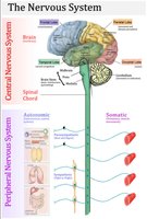
Anatomical Subdivisions of the Nervous System
Central Nervous System (CNS) and Peripheral Nervous System (PNS)
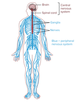
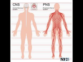
The nervous system is divided into two main anatomical subdivisions:
Central Nervous System (CNS): Consists of the brain and spinal cord. It acts as the integrating and control center, processing information and coordinating responses.
Peripheral Nervous System (PNS): Composed of cranial nerves (12 pairs), spinal nerves (31 pairs), and their branches. It connects the CNS to limbs and organs, serving as a communication relay.
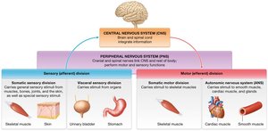
Functional Subdivisions of the PNS
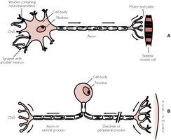
Sensory (Afferent) Division: Transmits sensory information from receptors to the CNS.
Motor (Efferent) Division: Transmits commands from the CNS to effector organs (muscles and glands).
Somatic Nervous System: Controls voluntary movements via skeletal muscles.
Autonomic Nervous System (ANS): Regulates involuntary functions (smooth muscle, cardiac muscle, glands) and is further divided into sympathetic and parasympathetic divisions.
Major Functions of the Nervous System
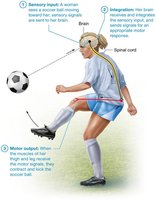
Sensory Input, Integration, and Motor Output
The nervous system performs three primary functions:
Sensory Input: Gathering information from sensory receptors about internal and external changes.
Integration: Processing and interpreting sensory input to determine an appropriate response.
Motor Output: Activating effector organs (muscles and glands) to produce a response.
Nervous Tissue: Structure and Components
Cellular Composition of Nervous Tissue
Nervous tissue is specialized for communication and consists of two main cell types:
Neurons: Excitable cells responsible for transmitting electrical signals. They make up about 10% of nervous tissue cells.
Neuroglia (Glial Cells): Supportive cells that maintain the environment, assist neural function, and repair tissue. They constitute about 90% of nervous tissue cells.
Extracellular Matrix (ECM): Composed of ground substance and glycoproteins, making up about 20% of tissue volume.
Structure of a Typical Neuron
Neurons are highly specialized, long-lived, and mostly amitotic cells. Their structure supports rapid and directed communication:
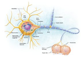
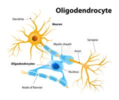
Cell Body (Soma): Contains the nucleus, cytoplasm, mitochondria, Nissl bodies (ribosomes and rough ER), and Golgi apparatus. Responsible for metabolic maintenance and protein synthesis.
Dendrites: Short, branched processes that receive signals and generate local potentials.
Axon: Long process that conducts action potentials away from the cell body. Features include the axon hillock (trigger zone), axon collaterals (branches), telodendria (terminal branches), and synaptic knobs (release neurotransmitters).
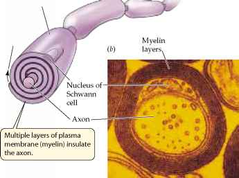
Myelin Sheath: Insulating layer that increases signal transmission speed.
Neuron Processes and Myelination
Axonal Transport: Movement of materials along the axon using motor proteins (kinesin for anterograde, dynein for retrograde transport).
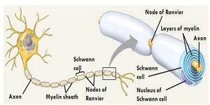
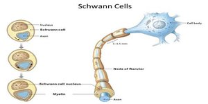
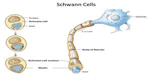
Myelin Sheath: Formed by Schwann cells in the PNS and oligodendrocytes in the CNS. Acts as an electrical insulator and increases conduction velocity.
Nodes of Ranvier: Gaps between myelin segments that facilitate rapid signal transmission (saltatory conduction).
Classification of Neurons
Structural Classification
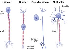
Multipolar Neurons: One axon, many dendrites; most common type, especially in CNS.
Bipolar Neurons: One axon, one dendrite; rare, found in the retina.
Unipolar (Pseudounipolar) Neurons: Single process that splits into two branches; primarily sensory neurons in the PNS.
Functional Classification
Sensory (Afferent) Neurons: Transmit impulses from receptors to the CNS; mostly unipolar.
Motor (Efferent) Neurons: Carry impulses from the CNS to effectors; cell bodies in the CNS.
Interneurons: Connect sensory and motor neurons; primarily multipolar and located in the CNS.
Neuroglia: Support Cells of the Nervous System
Types and Functions of Neuroglia
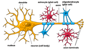
Neuroglia are essential for the maintenance, protection, and support of neurons. There are six main types:
CNS Neuroglia:
Astrocytes: Anchor neurons, maintain extracellular environment, form blood-brain barrier, repair tissue.
Oligodendrocytes: Form myelin sheaths around CNS axons.
Microglial Cells: Act as phagocytes, removing debris and pathogens.
Ependymal Cells: Line brain ventricles, produce and circulate cerebrospinal fluid.
PNS Neuroglia:
Schwann Cells: Form myelin sheaths around PNS axons.
Satellite Cells: Surround and support neuron cell bodies in ganglia.
Myelination in the CNS and PNS
Comparison of Myelination
CNS (Oligodendrocytes): Each cell can myelinate multiple axons; no neurolemma; myelination wraps from outside in.
PNS (Schwann Cells): Each cell myelinates a single axon segment; outer layer forms neurolemma; myelination wraps from inside out.
White Matter: Composed of myelinated axons; appears white.
Gray Matter: Composed of neuron cell bodies, unmyelinated dendrites, and axons; appears gray.
Regeneration and Repair in the Nervous System
Axon Repair and Regeneration
PNS: Axon regeneration is possible if the cell body and part of the myelinated axon remain. Schwann cells form a regeneration tube to guide axon regrowth.
CNS: Astrocytes form scar tissue, preventing axon regeneration; damaged neurons cannot be replaced.
Wallerian Degeneration: Process by which the distal portion of a severed axon degenerates, followed by regrowth from the proximal end.
Additional info: Neurons are amitotic after development due to loss of centrioles, making CNS repair especially limited.
