 Indietro
Nervous Tissue and Physiology II: Neural Electrophysiology and Synaptic Transmission
Guida di studio - Note intelligenti
 Appunti personalizzati basati sui tuoi materiali, ampliati con definizioni chiave, esempi e contesto.
Nervous Tissue and Physiology II
Neural Electrophysiology Overview
This section explores the fundamental principles of neural electrophysiology, including the generation and propagation of membrane potentials, local and action potentials, and the mechanisms of synaptic transmission. Understanding these concepts is essential for grasping how neurons communicate and process information.
Membrane Potential
Principles of Electricity in Neurons
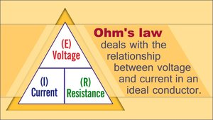
Neurons utilize electrical principles to transmit signals. The membrane potential is the voltage difference across the cell membrane, resulting from the separation of electrical charges.
Voltage (V): Measure of potential energy between two points; larger differences yield higher voltage.
Current (I): Flow of electrical charge from one point to another.
Resistance (R): Hindrance to the flow of electrical charge; insulators have high resistance, conductors have low resistance.
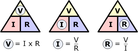
Ohm’s Law: Describes the relationship between voltage, current, and resistance:
Ion Channels and Membrane Potential
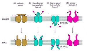
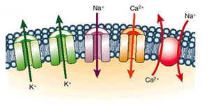
Ion channels are proteins that allow ions to move across the membrane, contributing to the membrane potential.
Chemically (Ligand) Gated: Open when a specific chemical binds.
Voltage Gated: Open in response to changes in membrane potential.
Mechanically Gated: Open by physical deformation.
Leak Channels: Always open, allowing passive ion movement.
Ions diffuse down their concentration and electrical gradients.
Resting Membrane Potential
The resting membrane potential is the difference in charge between the inside and outside of a neuron at rest, typically around -70 mV. It is maintained by the sodium-potassium pump and leak channels.
Na+/K+ Pump: Moves 3 Na+ out and 2 K+ in, creating a net negative charge inside.
Leak Channels: More K+ leak channels than Na+, allowing K+ to diffuse out more easily.
Polarized: Outside is positively charged, inside is negatively charged.
Ionic Composition: Cytosol has lower Na+ and higher K+; extracellular fluid has balanced cations and Cl-.
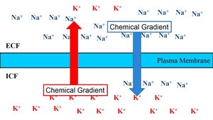
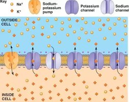
Changes in Membrane Potential
Membrane potential can change in response to stimuli:
Depolarization: Membrane potential becomes less negative (e.g., -70 mV to -65 mV).
Hyperpolarization: Membrane potential becomes more negative (e.g., -70 mV to -75 mV).
Graded Potential: Short-distance, variable changes in membrane potential.
Action Potential: Long-distance, all-or-nothing signal.
Local Potentials
Generation and Properties
Local potentials are small changes in membrane potential, typically generated by ligand-gated ion channels in dendrites and the cell body.
Graded Depolarization: Sodium ions enter, making the cell less negative.
Graded Hyperpolarization: Chloride ions enter, making the cell more negative.
Properties: Graded, reversible, decremental (decrease with distance).
Useful for short-distance signaling.
Action Potentials
Generation and Phases
Action potentials are rapid, long-distance electrical signals generated in excitable cells like neurons and muscle cells. They involve a reversal of membrane potential from -70 mV to +30 mV.
Resting State: All gated Na+ and K+ channels are closed.
Depolarization: Na+ channels open, threshold reached (~ -55 mV), positive feedback loop.
Repolarization: Na+ channels inactivate, K+ channels open.
Hyperpolarization: K+ channels remain open, Na+ channels reset.
Na+/K+ Pump: Restores ion distribution.
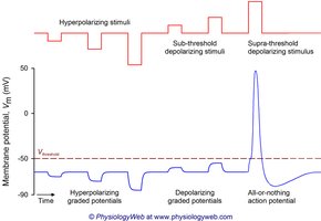
Threshold and Propagation
Action potentials are all-or-nothing events. Once threshold is reached, the action potential propagates along the axon without decrement.
Frequency: Determines stimulus intensity.
Propagation: Spreads from point of origin.
Refractory Periods
Refractory periods ensure unidirectional propagation and limit the frequency of action potentials.
Absolute Refractory Period: Neuron cannot respond; Na+ channels are inactivated.
Relative Refractory Period: Elevated threshold; K+ channels still open.
Action Potential Propagation
Saltatory vs. Continuous Conduction
Action potentials propagate differently in myelinated and unmyelinated axons.
Saltatory Conduction: In myelinated axons, action potentials "jump" between nodes of Ranvier, increasing speed and efficiency.
Continuous Conduction: In unmyelinated axons, action potentials propagate along every section of the axon, resulting in slower conduction.
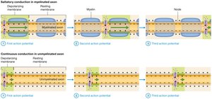
Conduction Velocity
Conduction velocity depends on axon diameter and degree of myelination.
Larger Diameter: Faster impulses due to less resistance.
More Myelin: Faster conduction via saltatory conduction.
Fiber Groups: Group A (fastest), Group B (intermediate), Group C (slowest).
Neuronal Synapses and Signal Transmission
Synapse Structure and Function
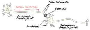
Synapses are junctions that mediate information transfer between neurons. They can be axodendritic or axosomatic.
Presynaptic Neuron: Transmits the signal.
Postsynaptic Neuron: Receives the signal.
Electrical Synapses
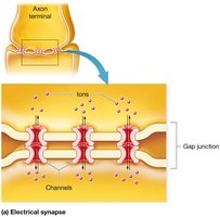
Electrical synapses involve gap junctions that allow ions to flow directly between cells, enabling rapid and coordinated activity.
Gap Junctions: Channels align to form pores for ion flow.
Location: Found in areas responsible for automatic behaviors and in cardiac/visceral smooth muscle.
Chemical Synapses
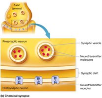
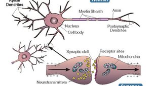
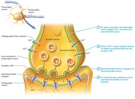
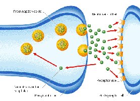
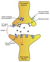
Chemical synapses are the most common type in the nervous system. They involve the release of neurotransmitters from synaptic vesicles into the synaptic cleft.
Synaptic Vesicles: Contain neurotransmitters.
Synaptic Cleft: Small space separating presynaptic and postsynaptic neurons.
Neurotransmitter Receptors: Bind neurotransmitters to initiate postsynaptic responses.
Events at Chemical Synapse
Synaptic transmission involves several steps:
Action potential arrives at axon terminal.
Voltage-gated Ca2+ channels open; Ca2+ enters axon terminal.
Ca2+ influx triggers neurotransmitter release via exocytosis.
Neurotransmitter diffuses across synaptic cleft and binds to receptors.
Binding opens ion channels, creating a graded potential.
Neurotransmitter effects are terminated by reuptake, degradation, or diffusion.
Excitation and Inhibition
Postsynaptic Potentials
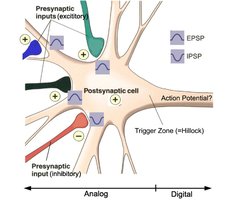
Postsynaptic potentials are local changes in membrane potential in the postsynaptic neuron.
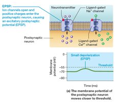
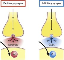
Excitatory Postsynaptic Potential (EPSP): Small local depolarization; moves membrane potential closer to threshold.
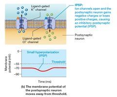
Inhibitory Postsynaptic Potential (IPSP): Small local hyperpolarization; moves membrane potential farther from threshold.
Termination of Synaptic Transmission
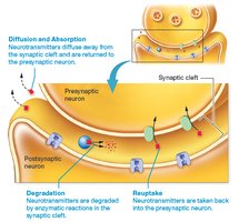
Neurotransmitter effects are terminated by:
Reuptake: Neurotransmitter is taken back into the presynaptic neuron.
Degradation: Enzymes break down neurotransmitter.
Diffusion: Neurotransmitter moves away from synaptic cleft.
Integration and Summation
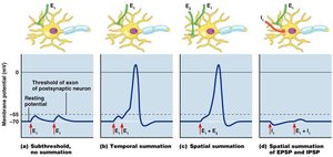
Neurons integrate multiple inputs through summation:
Temporal Summation: Rapid impulses from presynaptic neurons.
Spatial Summation: Large stimulation events with much neurotransmitter released.
Facilitated Neurons: Partially depolarized, more easily excited.
Synaptic Potentiation: Repeated use enhances synapse's ability to be excited.
Presynaptic Inhibition: One neuron inhibits another's impulse.
Neurotransmitters and Receptors Classification
Neurotransmitter Function and Types
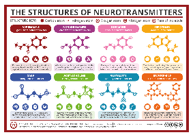
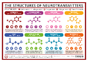
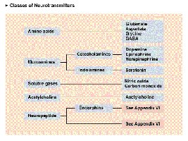
Neurotransmitters are chemicals released by neurons to transmit signals across synapses. They can be classified by function and chemical structure.
Excitatory: Cause depolarization (e.g., glutamate).
Inhibitory: Cause hyperpolarization (e.g., GABA, glycine).
Direct: Open ion channels directly.
Indirect: Activate secondary messenger systems.
Neuromodulators: Affect strength of transmission.
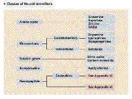
Major Neurotransmitter Classes
Acetylcholine (ACh): Stimulates skeletal muscles, found in ANS.
Biogenic Amines: Includes catecholamines (dopamine, norepinephrine, epinephrine) and indolamines (serotonin, histamine).
Amino Acids: Glutamate, aspartate, glycine, GABA.
Peptides: Substance P (pain), endorphins (pain relief), somatostatin, CCK.
Purines: ATP, adenosine.
Gases and Lipids: Nitric oxide, carbon monoxide, hydrogen sulfide, endocannabinoids.
Complex Behaviors and Neural Circuits
Neuronal Pools and Processing
Neuronal pools are groups of neurons that process information collectively. Neural processing can be serial (step-by-step, reflexes) or parallel (simultaneous input to different pathways).
Reflex Arcs: Pathways for automatic responses.
Types of Circuits: Divergent, convergent, reverberating, parallel after-discharge.
*Additional info: Expanded explanations and context were added to ensure completeness and academic quality, including definitions, examples, and mechanisms for each topic.*
