 Indietro
Nervous Tissue and Physiology: Structure and Function
Guida di studio - Note intelligenti
 Appunti personalizzati basati sui tuoi materiali, ampliati con definizioni chiave, esempi e contesto.
Nervous System Overview
Organs and Structure of the Nervous System
The human nervous system is a complex network that enables communication, environmental response, and regulation of bodily functions. It consists of specialized organs and tissues that work together to transmit signals throughout the body.
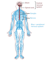
Brain: The central organ responsible for processing information and controlling responses.
Spinal Cord: Connects the brain to peripheral nerves and mediates reflexes.
Nerves: Bundles of axons that transmit signals between the CNS and the rest of the body.
Ganglia: Collections of neuron cell bodies in the PNS.
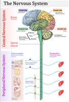
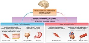
Anatomical Subdivisions: CNS and PNS
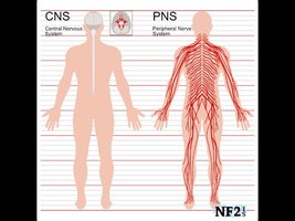
The nervous system is divided into two main anatomical subdivisions:
Central Nervous System (CNS): Includes the brain and spinal cord. Acts as the integrating and control center.
Peripheral Nervous System (PNS): Composed of cranial nerves, spinal nerves, and their branches. Connects the CNS to limbs and organs.
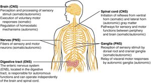
Functional Subdivisions of the Nervous System
Three Primary Functions
The nervous system performs three essential functions:
Sensory Input: Gathering information from internal and external environments.
Integration: Processing and interpreting sensory input to determine appropriate responses.
Motor Output: Activating effector organs (muscles and glands) to produce a response.
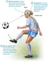
Functional Subdivisions: Somatic and Autonomic
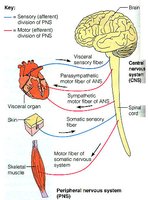
The PNS is further divided into functional subdivisions:
Somatic Nervous System: Controls voluntary movements via skeletal muscles.
Autonomic Nervous System (ANS): Regulates involuntary functions such as heart rate, digestion, and glandular activity. Includes sympathetic and parasympathetic divisions.
Nervous Tissue Composition
Cellular Components
Nervous tissue is primarily composed of two cell types:
Neurons: Excitable cells responsible for transmitting electrical signals. Make up about 10% of nervous tissue cells.
Neuroglia (Glial Cells): Supportive cells that maintain the environment, assist neural function, and repair tissue. Make up about 90% of nervous tissue cells.
Extracellular Matrix (ECM)
Composed of ground substance and glycoproteins.
Provides structural support and regulates the environment for neurons and glia.
Neuron Structure and Function
Parts of a Typical Neuron
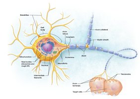
Neurons are specialized for rapid communication and consist of several distinct regions:
Cell Body (Soma): Contains the nucleus, cytoplasm, and organelles. Responsible for metabolic activities and protein synthesis.
Dendrites: Short, branched processes that receive signals from other neurons or sensory receptors.
Axon: Long process that conducts action potentials away from the cell body. May be wrapped in a myelin sheath for insulation and increased transmission speed.
Axon Hillock: Junction where action potentials are initiated.
Axon Collaterals: Branches off the main axon.
Telodendria: Terminal branches ending in synaptic knobs.
Myelin Sheath and Nodes of Ranvier
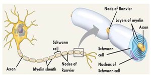
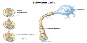
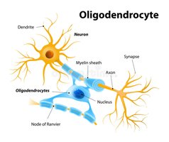
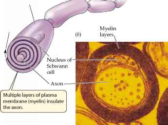
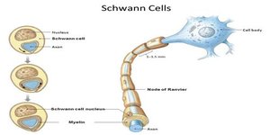
Myelin Sheath: A multilayered lipid and protein covering produced by glial cells. Acts as an electrical insulator and increases the speed of nerve impulse transmission.
Nodes of Ranvier: Gaps between myelin segments where action potentials are regenerated.
Axonal Transport
Axonal transport is essential for moving materials between the cell body and axon terminals.
Slow Axonal Transport: Moves cytoskeletal and other proteins anterogradely at 1-3 mm/day.
Fast Axonal Transport: Moves vesicles and organelles both anterogradely and retrogradely at 200-400 mm/day.
Neuron Classification
Structural Classification
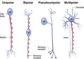
Neurons are classified based on the number and arrangement of their processes:
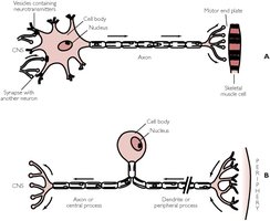
Multipolar: One axon, multiple dendrites. Most common type.
Bipolar: One axon, one dendrite. Rare, found in the retina.
Unipolar (Pseudounipolar): Single process that splits into two branches. Found primarily in sensory ganglia of the PNS.
Functional Classification
Sensory (Afferent) Neurons: Transmit impulses from receptors to the CNS. Mostly unipolar.
Motor (Efferent) Neurons: Carry impulses away from the CNS to effectors. Cell bodies located in the CNS.
Interneurons: Connect sensory and motor neurons. Primarily multipolar and confined to the CNS.
Neuroglia (Glial Cells)
Types and Functions
Neuroglia are essential for supporting neurons and maintaining nervous tissue health.
CNS Neuroglia:
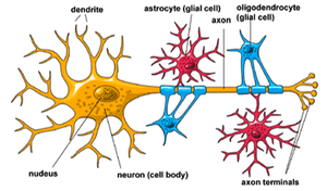
Astrocytes: Anchor neurons, maintain extracellular environment, assist in blood-brain barrier formation, and repair tissue.
Oligodendrocytes: Form myelin sheaths around CNS axons.
Microglial Cells: Act as macrophages, phagocytizing foreign invaders and debris.
Ependymal Cells: Line brain ventricles, produce and circulate cerebrospinal fluid.
PNS Neuroglia:
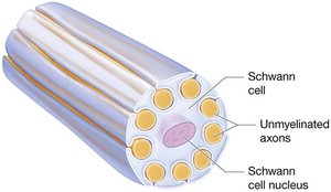
Schwann Cells: Form myelin sheaths around PNS axons.
Satellite Cells: Surround and support neuron cell bodies in ganglia.
Myelination in CNS and PNS
Differences in Myelination
CNS: Oligodendrocytes wrap around multiple axons. Myelination begins after birth. No neurolemma.
PNS: Schwann cells wrap around a single axon. Myelination begins in early fetal period. Neurolemma present.
Axon Repair and Regeneration
Repair Mechanisms
PNS: Axon regeneration is possible if the cell body and part of the myelinated axon remain. Schwann cells and basal lamina form a regeneration tube to guide regrowth.
CNS: Astrocytes form scar tissue, preventing neuron replacement.
Summary Table: CNS vs. PNS
Feature | CNS | PNS |
|---|---|---|
Main Structures | Brain, Spinal Cord | Nerves, Ganglia |
Myelinating Cells | Oligodendrocytes | Schwann Cells |
Repair Ability | Limited (scar tissue) | Possible (regeneration tube) |
Functional Divisions | Integration, Control | Sensory, Motor, Autonomic |
Key Terms and Concepts
Neuron: Excitable cell transmitting electrical signals.
Neuroglia: Supportive cells in nervous tissue.
Myelin Sheath: Insulating layer around axons.
Nodes of Ranvier: Gaps in myelin sheath for signal regeneration.
Axonal Transport: Movement of materials along axon.
Ganglia: Clusters of neuron cell bodies in PNS.
Nuclei: Clusters of neuron cell bodies in CNS.
Equations and Formulas
Resting Membrane Potential
The resting membrane potential of neurons is determined by ion concentrations and membrane permeability.
Typical value:
Formula:
Action Potential Propagation
Speed increases with myelination and axon diameter.
Saltatory conduction occurs at nodes of Ranvier.
Example: Reflex Arc
A sensory neuron detects heat and sends an afferent signal to the CNS.
The CNS integrates the signal and sends an efferent signal via a motor neuron to contract muscles and withdraw the finger.
Additional info:
Neurons are amitotic and long-lived, requiring high metabolic rates.
Rabies, poliovirus, and tetanus toxin use retrograde axonal transport to infect the CNS.
