 Indietro
Nervous Tissue and Physiology: Structure, Function, and Cellular Organization
Guida di studio - Note intelligenti
 Appunti personalizzati basati sui tuoi materiali, ampliati con definizioni chiave, esempi e contesto.
Chapter 12: Nervous Tissue and Physiology
Case Study: Neurological Symptoms and Diagnostic Tools
This case study illustrates the complexity of neurological symptoms and the importance of diagnostic imaging in identifying underlying causes. MRI (Magnetic Resonance Imaging) and EEG (Electroencephalogram) are recommended to detect structural and functional abnormalities in the nervous system.
MRI: Provides detailed images of brain and spinal cord structures, useful for identifying lesions, demyelination, or tumors.
EEG: Measures electrical activity in the brain, helpful for diagnosing epilepsy, abnormal brain function, or other neurological disorders.
Example: Multiple sclerosis may present with symptoms such as muscle cramps, vision changes, and abnormal sensory perceptions, which can be detected by MRI and EEG.
Functions of the Nervous System
Main Functions
The nervous system is responsible for receiving sensory input, integrating information, controlling muscles and glands, maintaining homeostasis, and establishing mental activity.
Receiving sensory input: Detects internal and external stimuli (e.g., touch, temperature).
Integrating information: Processes sensory input and initiates responses (e.g., reflexes).
Controlling muscles and glands: Initiates movement and regulates glandular activity.
Maintaining homeostasis: Coordinates physiological processes (e.g., heart rate, breathing).
Establishing and maintaining mental activity: Supports consciousness, thinking, memory, and emotion.
Example: Reflex withdrawal from a hot object involves sensory input, integration, and motor output.
Divisions of the Nervous System
Central and Peripheral Nervous Systems
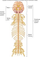
The nervous system is divided into the Central Nervous System (CNS) and Peripheral Nervous System (PNS).
CNS: Consists of the brain and spinal cord; responsible for processing and integrating information.
PNS: Composed of sensory receptors, nerves, ganglia, and plexuses; connects CNS to the rest of the body.
Components of the PNS
Sensory receptors: Specialized cells that detect stimuli (e.g., temperature, pain).
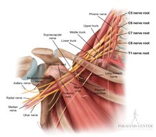
Nerves: Bundles of axons connecting CNS to sensory receptors, muscles, and glands.
Cranial nerves: Originate from the brain (12 pairs).
Spinal nerves: Originate from the spinal cord (31 pairs).
Ganglia: Collections of neuron cell bodies outside the CNS.
Plexus: Networks of axons and sometimes neuron cell bodies outside the CNS.
Divisions of the PNS
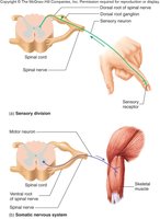
Sensory (afferent) division: Transmits action potentials from receptors to CNS.
Motor (efferent) division: Transmits action potentials from CNS to effectors (muscles, glands).
Effectors: Target organs or tissues (muscles, glands) that respond to neural commands.
Somatic and Autonomic Nervous Systems
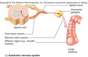
Somatic nervous system: Controls voluntary movements via skeletal muscles; single neuron system.
Autonomic nervous system (ANS): Controls involuntary functions (smooth muscle, cardiac muscle, glands); two-neuron system.
Divisions of the ANS
Sympathetic division: Prepares the body for physical activity (fight or flight response).
Parasympathetic division: Regulates resting functions (digestion, bladder control).
Enteric division: Controls digestive tract independently but interacts with sympathetic and parasympathetic divisions.
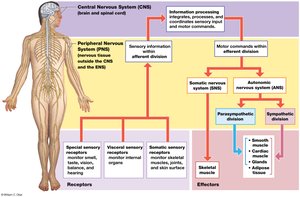
Organization of the Nervous System
Pathway from Receptor to Effector
The nervous system processes information through a pathway: receptor → sensory nervous system → CNS → motor nervous system → effector.
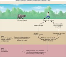
Cells of the Nervous System
Neurons and Neuroglia
Nervous tissue consists of neurons (nerve cells) and neuroglia (supporting cells).
Neurons: Receive stimuli and transmit action potentials; composed of cell body (soma), dendrites (input), and axons (output).
Neuroglia: Support and protect neurons; various types in CNS and PNS.
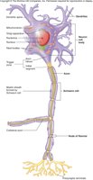
Parts of the Neuron
Cell body (soma): Contains nucleus and Nissl substance (site of protein synthesis).
Dendrites: Highly branched, receive input from other neurons.
Axon: Conducts action potentials; may branch to form collaterals.
Axon hillock: Initial segment where action potentials are generated.
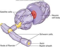
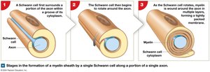
Schwann cells: Form myelin sheath around axons in PNS.
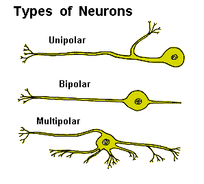
Types of Neurons
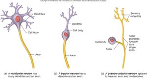
Multipolar: Many dendrites, one axon (most common in CNS).
Bipolar: One dendrite, one axon (sensory organs).
Pseudo-unipolar: Single process that splits into two branches (sensory neurons).
Unipolar: One process from cell body (rare in humans).
Neuroglia: Types and Functions
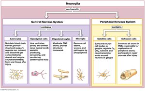
Neuroglia of the CNS
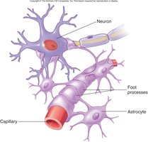
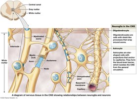
Astrocytes: Form blood-brain barrier, regulate extracellular fluid, provide structural support.
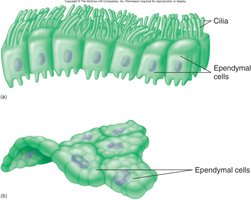
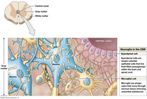
Ependymal cells: Line ventricles and central canal, secrete cerebrospinal fluid (CSF).
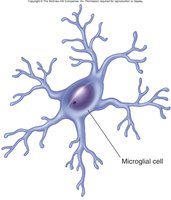
Microglia: Specialized macrophages, respond to inflammation, phagocytize debris.
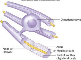
Oligodendrocytes: Form myelin sheaths around CNS axons.
Neuroglia of the PNS
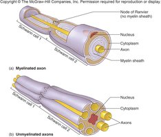
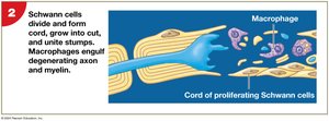
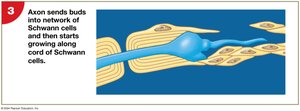
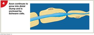
Schwann cells: Form myelin sheath around a single axon; aid in regeneration after injury.
Satellite cells: Surround neuron cell bodies in ganglia, provide support and nutrients.
Summary Table: Neuroglia in CNS and PNS
Neuroglia Type | Location | Function |
|---|---|---|
Astrocytes | CNS | Maintain blood-brain barrier, structural support |
Ependymal cells | CNS | Produce and circulate CSF |
Oligodendrocytes | CNS | Form myelin sheaths |
Microglia | CNS | Phagocytosis, immune defense |
Schwann cells | PNS | Form myelin sheaths, aid regeneration |
Satellite cells | PNS | Support neuron cell bodies |
Neural Responses to Injury and Demyelination
Demyelination
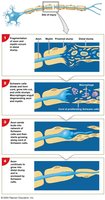
Demyelination: Progressive destruction of myelin sheaths in CNS or PNS.
Causes: Heavy-metal exposure, diphtheria, multiple sclerosis (MS).
Effects: Impaired nerve conduction, motor and sensory deficits.
Peripheral Nerve Regeneration
Wallerian degeneration: Axon distal to injury degenerates; Schwann cells proliferate and guide regrowth.
CNS regeneration: Limited by astrocyte scar tissue and inhibitory chemicals.
Resting Membrane Potential (RMP) and Ion Channels
Resting Membrane Potential
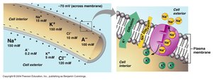
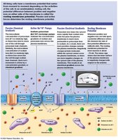
The resting membrane potential is the electrical charge difference across the plasma membrane of a neuron, typically around -70 mV.
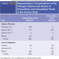
Key factors: Membrane permeability to ions, concentration gradients, and activity of the Na+/K+ pump.
Equation: (Nernst equation for potassium)
Example: Potassium (K+) is more concentrated inside the cell, sodium (Na+) outside.
Types of Ion Channels
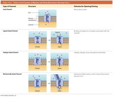
Leak channels: Always open, allow ions to flow down concentration gradients.
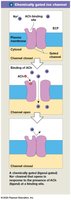
Gated channels: Open in response to specific stimuli.
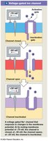
Voltage-gated channels: Open in response to voltage changes.
Ligand-gated channels: Open in response to ligand binding.
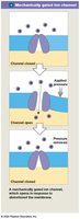
Mechanically-gated channels: Open in response to mechanical stimulation.
Graded Potentials and Action Potentials
Graded Potentials
Graded potentials are local changes in membrane potential that vary in magnitude and can summate to trigger action potentials.
Causes: Ligand binding, mechanical stimulation, temperature changes.
Spread: Decremental, decrease in magnitude as they move across the membrane.
Can trigger: Action potentials if threshold is reached.
Action Potentials
Action potentials are rapid, uniform depolarization and repolarization events that propagate along axons.
Depolarization: Membrane becomes more positive due to Na+ influx.
Repolarization: Membrane returns to negative due to K+ efflux.
All-or-none principle: Once threshold is reached, action potential occurs regardless of stimulus strength.
Threshold: Minimum membrane potential required to trigger an action potential.
Sodium and Potassium Channels in Action Potentials
Voltage-gated sodium channels: Have activation and inactivation gates; three states (resting, activated, inactivated).
Voltage-gated potassium channels: Two states (resting, activated).
Summary Table: Ion Effects on Resting Membrane Potential
Ion | Effect on RMP |
|---|---|
Na+ | Moves in, depolarizes membrane |
K+ | Moves out, hyperpolarizes membrane |
Ca2+ | Moves in, depolarizes membrane |
Cl- | Moves in, hyperpolarizes membrane |
Additional info:
Potassium deficiency can cause symptoms resembling a heart attack due to its role in maintaining membrane potential and proper muscle contraction, including cardiac muscle.
Resting membrane potential is maintained by the Na+/K+ pump and selective permeability of the membrane to different ions.
