 Indietro
Nervous Tissue and the Nervous System: Structure, Function, and Physiology
Guida di studio - Note intelligenti
 Appunti personalizzati basati sui tuoi materiali, ampliati con definizioni chiave, esempi e contesto.
Nervous System: Anatomical and Functional Divisions
Overview of the Nervous System
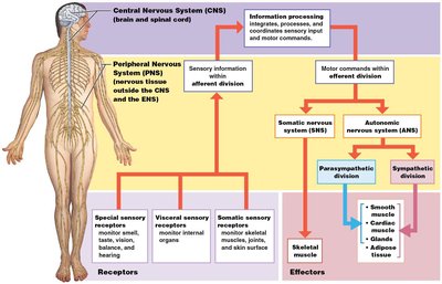
The nervous system is a complex network responsible for communication, information processing, and control throughout the body. It consists of the central nervous system (CNS), peripheral nervous system (PNS), and enteric nervous system (ENS), each with distinct roles and components.
Central Nervous System (CNS): Includes the brain and spinal cord; responsible for information processing, integration, and coordination of sensory input and motor commands.
Peripheral Nervous System (PNS): Composed of nerves and ganglia outside the CNS; divided into afferent (sensory) and efferent (motor) divisions.
Somatic Nervous System (SNS): Controls voluntary and involuntary skeletal muscle contractions.
Autonomic Nervous System (ANS): Regulates involuntary functions, including smooth and cardiac muscle activity and glandular secretions; subdivided into sympathetic (stimulating) and parasympathetic (relaxing) divisions.
Enteric Nervous System (ENS): Governs the function of the gastrointestinal tract independently, but can be influenced by the ANS.
Neurons: Structure and Function
Anatomy of a Typical Neuron
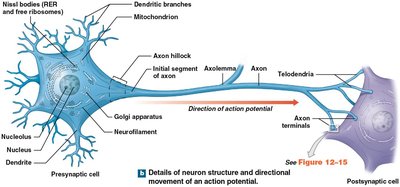
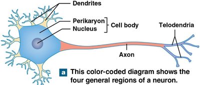
Neurons are the basic functional units of the nervous system, specialized for communication and information processing. Each neuron consists of distinct regions that contribute to its function.
Cell Body (Soma): Contains the nucleus and organelles; site of metabolic activity.
Dendrites: Receive incoming signals from other neurons.
Axon: Conducts electrical impulses away from the cell body.
Axon Terminals: Transmit signals to other cells via synapses.
Telodendria: Fine extensions at the end of the axon.
Structural Classification of Neurons
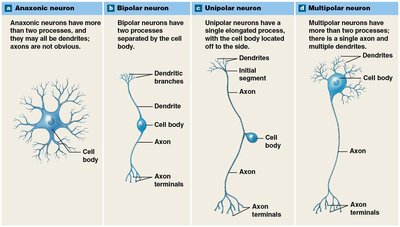
Neurons are classified based on their structure, which relates to their function and location in the nervous system.
Anaxonic Neurons: Small, indistinguishable axons and dendrites; found in the brain and special sense organs.
Bipolar Neurons: One dendrite and one axon; rare, found in special sense organs (e.g., retina).
Unipolar Neurons: Fused axon and dendrite; cell body to one side; most sensory neurons in the PNS.
Multipolar Neurons: One long axon and multiple dendrites; common in CNS; all motor neurons controlling skeletal muscles.
Functional Classification of Neurons
Sensory Neurons: Somatic sensory neurons monitor external environment; visceral sensory neurons monitor internal environment.
Motor Neurons: Efferent neurons carry instructions from CNS to effectors; somatic motor neurons innervate skeletal muscles; visceral motor neurons innervate smooth and cardiac muscle, glands, adipose tissue.
Interneurons: Located in CNS; distribute sensory information and coordinate motor activity; involved in higher functions such as memory, planning, and learning.
Neuroglia: Support Cells of the Nervous System
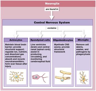
Neuroglia in the Central Nervous System
Neuroglia are essential for the survival and function of neurons, providing structural and metabolic support. They make up half the volume of the nervous system.
Astrocytes: Maintain blood-brain barrier, provide structural support, regulate ion and nutrient concentrations, absorb/recycle neurotransmitters, form scar tissue after injury.
Ependymal Cells: Line ventricles and central canal; produce, circulate, and monitor cerebrospinal fluid.
Oligodendrocytes: Myelinate CNS axons; provide structural framework.
Microglia: Remove cell debris, waste, and pathogens by phagocytosis.
Neuroglia in the Peripheral Nervous System
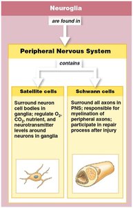
Satellite Cells: Surround neuron cell bodies in ganglia; regulate O2, CO2, nutrient, and neurotransmitter levels.
Schwann Cells: Surround all axons in PNS; responsible for myelination and repair after injury.
Membrane Potential and Electrical Signaling
Resting Membrane Potential
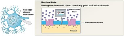
The resting membrane potential is the electrical charge difference across the plasma membrane, maintained by ion gradients and selective permeability. It is crucial for neuronal function.
Typical resting membrane potential: −70 mV
Key ions: Sodium (Na+), Potassium (K+)
Sodium–potassium pump: Ejects 3 Na+ for every 2 K+ brought in, powered by ATP
Equilibrium potentials: K+ = −90 mV, Na+ = +66 mV
Graded Potentials
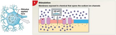
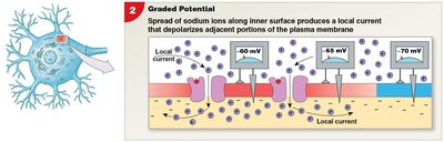
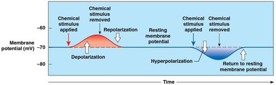
Graded potentials are local changes in membrane potential that occur in response to stimuli. They can lead to depolarization or hyperpolarization and are most pronounced at the site of stimulation.
Depolarization: Membrane potential becomes less negative (closer to zero).
Hyperpolarization: Membrane potential becomes more negative.
Repolarization: Return to resting membrane potential after stimulus removal.
Characteristics: Effect decreases with distance; stronger stimuli produce greater changes.
Action Potentials: Generation and Propagation
Generation of an Action Potential
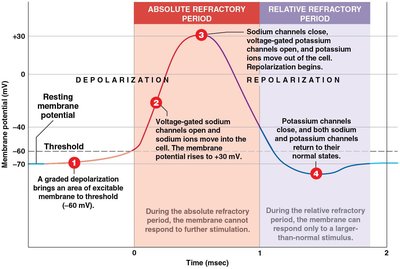
An action potential is a rapid, large change in membrane potential that propagates along the axon, enabling communication between neurons.
Threshold: A graded depolarization brings the membrane to threshold (about −60 mV).
Depolarization: Voltage-gated sodium channels open; Na+ enters cell; membrane potential rises to +30 mV.
Repolarization: Sodium channels close; potassium channels open; K+ leaves cell; membrane potential returns to resting state.
Refractory Periods: Absolute (no further action potential possible) and relative (stronger stimulus required).
Propagation of Action Potentials
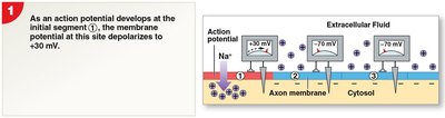
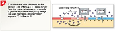
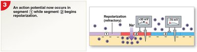
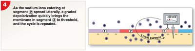
Action potentials propagate along unmyelinated axons by continuous conduction, affecting one segment at a time. The cycle repeats, ensuring one-way transmission.
Step 1: Action potential develops at initial segment; depolarizes to +30 mV.
Step 2: Local current develops; depolarizes next segment to threshold.
Step 3: Action potential occurs in second segment; initial segment begins repolarization.
Step 4: Local current depolarizes next segment; cycle repeats.
Synaptic Activity and Neurotransmitters
Structure and Function of Synapses
A synapse is a specialized site where a neuron communicates with another cell. The presynaptic neuron sends the message, and the postsynaptic neuron receives it.
Electrical Synapses: Direct physical contact between cells.
Chemical Synapses: Signal transmitted across a gap by neurotransmitters.
Neurotransmitters
Neurotransmitters are chemical messengers released from synaptic vesicles in the axon terminal. They affect receptors on the postsynaptic membrane, are broken down by enzymes, and reabsorbed for reuse.
Cholinergic: Acetylcholine (ACh)
Biogenic amines: Nor-epinephrine (Noradrenaline), Dopamine, Serotonin
Amino acids: GABA
Neuropeptides: Opioids
Dissolved gases: Nitric oxide, Carbon monoxide
Summary Table: Structural Classification of Neurons
Type | Structure | Location |
|---|---|---|
Anaxonic | Indistinguishable axons/dendrites | Brain, special sense organs |
Bipolar | One dendrite, one axon | Special sense organs |
Unipolar | Fused axon/dendrite, cell body to side | Sensory neurons of PNS |
Multipolar | One axon, multiple dendrites | CNS, motor neurons |
Summary Table: Neuroglia Types and Functions
System | Cell Type | Function |
|---|---|---|
CNS | Astrocytes | Support, blood-brain barrier, neurotransmitter recycling |
CNS | Ependymal cells | Produce/monitor cerebrospinal fluid |
CNS | Oligodendrocytes | Myelinate axons, structural framework |
CNS | Microglia | Phagocytosis of debris/pathogens |
PNS | Satellite cells | Regulate environment around neurons |
PNS | Schwann cells | Myelinate axons, repair after injury |
Key Equations
Sodium–Potassium Pump:
Resting Membrane Potential:
Equilibrium Potential for K+:
Equilibrium Potential for Na+:
Additional info: Academic context was added to clarify the functional and structural roles of neurons and neuroglia, as well as the physiological basis of membrane potentials and action potentials.
