 Indietro
Overview of the Nervous System: Structure and Function
Guida di studio - Note intelligenti
 Appunti personalizzati basati sui tuoi materiali, ampliati con definizioni chiave, esempi e contesto.
The Nervous System: Structure and Organization
Central and Peripheral Nervous System
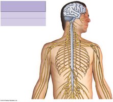
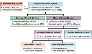
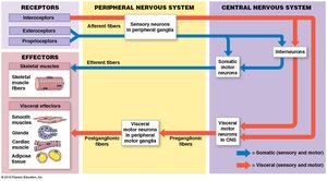
The nervous system is divided into two main anatomical and functional divisions: the Central Nervous System (CNS) and the Peripheral Nervous System (PNS). The CNS consists of the brain and spinal cord, serving as the integrative and control center. The PNS includes all neural elements outside the CNS, such as cranial and spinal nerves, and acts as a communication link between the CNS and the rest of the body.
Central Nervous System (CNS): Brain and spinal cord; processes and integrates information.
Peripheral Nervous System (PNS): Cranial and spinal nerves; transmits signals to and from the CNS.
Functional Divisions of the Nervous System
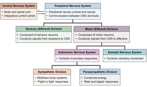
The nervous system is further divided functionally into sensory (afferent) and motor (efferent) divisions. The sensory division carries information from receptors to the CNS, while the motor division transmits commands from the CNS to effectors (muscles and glands).
Sensory (Afferent) Division: Composed of sensory neurons; conducts signals from receptors to the CNS.
Motor (Efferent) Division: Composed of motor neurons; conducts signals from the CNS to effectors.
Autonomic and Somatic Nervous Systems
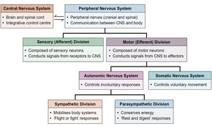
The motor division is subdivided into the Autonomic Nervous System (ANS) and the Somatic Nervous System. The ANS controls involuntary responses and is further divided into the sympathetic and parasympathetic divisions. The somatic nervous system controls voluntary movements.
Autonomic Nervous System (ANS): Regulates involuntary functions (e.g., heart rate, digestion).
Somatic Nervous System: Controls voluntary movements of skeletal muscles.
Sympathetic Division: Mobilizes body systems for "fight or flight" responses.
Parasympathetic Division: Conserves energy; promotes "rest and digest" responses.
Neural Pathways: Receptors and Effectors
Receptors and Effectors
Neural pathways begin with receptors that detect changes in the environment and end with effectors that carry out the body's responses. Receptors can be classified as interoceptors, exteroceptors, and proprioceptors. Effectors include skeletal muscles (somatic effectors) and smooth muscle, cardiac muscle, and glands (visceral effectors).
Receptors: Specialized cells or nerve endings that respond to stimuli.
Effectors: Muscles or glands that respond to neural commands.
Somatic Effectors: Skeletal muscles.
Visceral Effectors: Smooth muscle, cardiac muscle, glands.
Neurons: Structure and Function
Neuron Anatomy
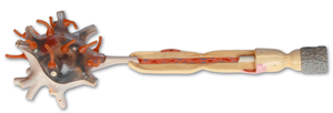
Neurons are the fundamental units of the nervous system, specialized for transmitting electrical impulses. Each neuron consists of a cell body (soma), dendrites, and an axon. Dendrites receive signals, while the axon transmits impulses away from the cell body.
Cell Body (Soma): Contains the nucleus and organelles.
Dendrites: Receive incoming signals.
Axon: Conducts electrical impulses to other neurons or effectors.
Types of Neurons
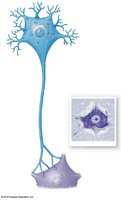
Neurons can be classified based on their function: sensory neurons, motor neurons, and interneurons. Sensory neurons transmit information from receptors to the CNS, motor neurons carry commands from the CNS to effectors, and interneurons connect neurons within the CNS.
Sensory Neurons: Carry information from receptors to the CNS.
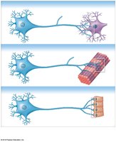
Motor Neurons: Transmit commands from the CNS to effectors.
Interneurons: Connect neurons within the CNS; involved in processing and integration.
Spinal Cord and Nerve Organization
Spinal Cord Structure
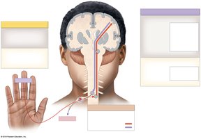
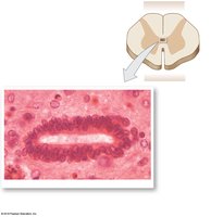
The spinal cord is a major pathway for information traveling between the brain and the rest of the body. It is organized into segments, each giving rise to a pair of spinal nerves. The spinal cord contains both gray matter (cell bodies) and white matter (myelinated axons).
Gray Matter: Contains neuron cell bodies; involved in processing information.
White Matter: Contains myelinated axons; responsible for transmitting signals.
Spinal Nerves and Dermatomes
Spinal nerves are mixed nerves that carry both sensory and motor fibers. Each spinal nerve innervates a specific region of the body, known as a dermatome. Dermatomes are important for clinical assessment of nerve function.
Spinal Nerves: Mixed nerves emerging from the spinal cord.
Dermatome: Area of skin supplied by a single spinal nerve.
Summary Table: Nervous System Divisions
The following table summarizes the main divisions and functions of the nervous system:
Division | Main Function | Key Structures |
|---|---|---|
Central Nervous System | Integration and processing | Brain, spinal cord |
Peripheral Nervous System | Communication | Cranial nerves, spinal nerves |
Sensory (Afferent) Division | Signal reception | Sensory neurons |
Motor (Efferent) Division | Signal transmission | Motor neurons |
Autonomic Nervous System | Involuntary control | Sympathetic, parasympathetic |
Somatic Nervous System | Voluntary control | Skeletal muscles |
Key Terms and Concepts
Neuron: Basic unit of the nervous system; transmits electrical impulses.
Synapse: Junction between two neurons or a neuron and an effector.
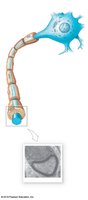
Myelin: Insulating layer around axons; increases speed of impulse transmission.
Reflex: Rapid, automatic response to a stimulus.
Example: Reflex Arc
A reflex arc is a simple neural pathway that mediates a reflex. It typically involves a sensory neuron, an interneuron, and a motor neuron.
Step 1: Receptor detects stimulus.
Step 2: Sensory neuron transmits signal to CNS.
Step 3: Interneuron processes information.
Step 4: Motor neuron transmits command to effector.
Step 5: Effector produces response.
Additional info:
Myelinated axons in white matter allow for rapid transmission of electrical impulses.
Gray matter is essential for synaptic integration and processing.
Clinical assessment of dermatomes helps diagnose nerve injuries.
