 Indietro
Parasympathetic Division of the Autonomic Nervous System: Structure, Function, and Mechanisms
Guida di studio - Note intelligenti
 Appunti personalizzati basati sui tuoi materiali, ampliati con definizioni chiave, esempi e contesto.
The Parasympathetic Division of the Autonomic Nervous System
Overview and Function
The parasympathetic division is one of two main branches of the autonomic nervous system (ANS), responsible for conserving energy and maintaining the body's resting metabolic rate. It is often described as the "rest and digest" system, in contrast to the sympathetic "fight or flight" response.
Primary Function: Promotes relaxation, energy conservation, and restoration of bodily resources.
Rest & Digest: Stimulates activities such as digestion, salivation, and reduced heart rate.
Metabolic Effects: Maintains homeostasis during periods of rest.

Anatomical Organization: Craniosacral Division
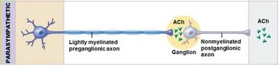
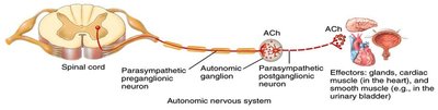
The parasympathetic division is also known as the craniosacral division due to its origin in the brainstem and sacral segments of the spinal cord. Its fibers are organized into long preganglionic and short postganglionic fibers.
Preganglionic Neurons: Located in the brainstem and lateral portion of the anterior gray horns of S2-S4 spinal segments.
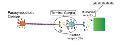
Ganglia: Located in or near target organs; preganglionic fibers release acetylcholine (ACh) to stimulate ganglionic neurons.
Postganglionic Fibers: Short, located within or near the target organ.
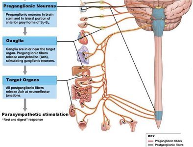
Parasympathetic Ganglia: Terminal and Intramural Ganglia
Parasympathetic ganglia are classified as terminal (located near target organs) or intramural (embedded within the walls of target organs). Preganglionic fibers exit the brain via cranial nerves III, VII, IX, and X, controlling various visceral structures.
Terminal Ganglia: Associated with cranial nerves III (oculomotor), VII (facial), IX (glossopharyngeal).
Intramural Ganglia: Located within the target organ, especially in the thoracic and abdominopelvic cavities.
Vagus Nerve (X): Provides 80% of all parasympathetic outflow, innervating neck, thoracic, and abdominopelvic structures.
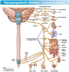
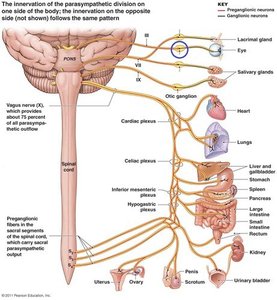
Parasympathetic Effects on Target Organs
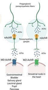
The effects of parasympathetic stimulation vary depending on the type of receptor and the nature of the second messenger involved in the target cell. All parasympathetic neurons (pre- and postganglionic) release acetylcholine (ACh).
Receptor Types: Effects depend on whether the postsynaptic cell expresses nicotinic or muscarinic receptors.
Second Messengers: The response may be excitatory or inhibitory based on enzyme activation or inactivation.
Examples of Effects: Pupil constriction, increased salivation, slowed heart rate, increased digestive activity.
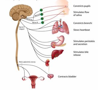
Neurotransmitter Release and Receptor Mechanisms
Acetylcholine (ACh) is the primary neurotransmitter released by both pre- and postganglionic parasympathetic neurons. The effects on target organs are mediated by two main receptor types: nicotinic and muscarinic.
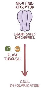
Nicotinic Receptors: Ligand-gated ion channels found on postganglionic neurons; activation leads to cell depolarization.
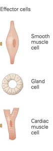
Muscarinic Receptors: G-protein coupled receptors found on effector cells; effects are longer lasting and can be excitatory or inhibitory.
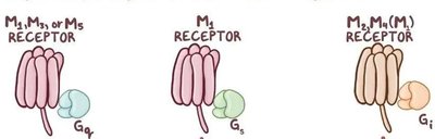
Muscarinic Receptor Subtypes and Their Effects
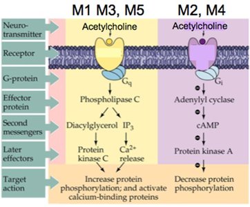
Muscarinic receptors are classified into five subtypes (M1–M5), each with distinct signaling pathways and effects on target organs.
M1, M3, M5: Stimulate phospholipase C (PLC) and increase intracellular Ca2+, leading to excitatory effects.
M2, M4: Inhibit adenylyl cyclase, decrease cAMP, and produce inhibitory effects.
Examples: M3 mediates smooth muscle contraction in the GI tract; M2 slows heart rate.
Parasympathetic Agonists and Antagonists
Drugs that mimic or block parasympathetic activity are called agonists and antagonists, respectively. These medications are used to treat various conditions by targeting cholinergic receptors.
Agonists: Methacholine, bethanechol—stimulate ACh receptors, used for urinary retention or tachycardia.
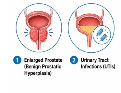
Antagonists: Atropine—blocks ACh receptors, used for overactive bladder and bradycardia.
Side Effects: Increased salivation, difficulty breathing, changes in pupil size, sweating.
Comparison of Sympathetic and Parasympathetic Divisions
The sympathetic and parasympathetic divisions differ in their anatomical organization, neurotransmitters, receptor types, and effects on target organs.
Feature | Sympathetic | Parasympathetic |
|---|---|---|
Origin | Thoracolumbar (T1–L2) | Craniosacral (brainstem, S2–S4) |
Ganglia Location | Near spinal cord | Near or within target organs |
Preganglionic Fiber Length | Short | Long |
Postganglionic Fiber Length | Long | Short |
Neurotransmitter (Postganglionic) | Norepinephrine (NE) | Acetylcholine (ACh) |
Receptor Types | Adrenergic | Muscarinic, Nicotinic |
Main Effects | Fight or flight | Rest and digest |
Summary of Key Points
The parasympathetic division is essential for maintaining homeostasis during rest.
It uses acetylcholine as its primary neurotransmitter, acting on nicotinic and muscarinic receptors.
Effects on target organs depend on receptor subtype and second messenger pathways.
Pharmacological agents can modulate parasympathetic activity for therapeutic purposes.
