 Indietro
Peripheral Nerve Regeneration and Neural Transmembrane Potential
Guida di studio - Note intelligenti
 Appunti personalizzati basati sui tuoi materiali, ampliati con definizioni chiave, esempi e contesto.
Peripheral Nerve Regeneration After Injury
Wallerian Degeneration
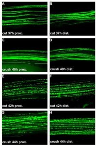
Wallerian degeneration is a process that occurs in peripheral nerves following injury, where the axon segment distal to the injury site degenerates. This phenomenon was first described by Augustus Waller in 1850. The degeneration typically begins within 24–36 hours after injury and is characterized by the breakdown of the axon and its myelin sheath.
Definition: Wallerian (anterograde) degeneration refers to the disintegration of the axon and myelin distal to the site of injury.
Timeline: Degeneration starts within 24–36 hours post-injury.
Example: Severing glossopharyngeal and hypoglossal nerves in experimental models.
Role of Schwann Cells in Peripheral Nerve Regeneration
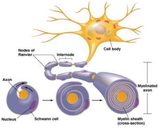
Schwann cells are essential for regeneration in the peripheral nervous system (PNS). They form a pathway for new axonal growth and wrap the regenerating axon in myelin. Unlike central nervous system (CNS) glial cells, Schwann cells maintain the neurolemma, which does not degenerate, facilitating regeneration.
Schwann Cells: Provide structural and biochemical support for axonal regrowth.
Myelination: Schwann cells wrap new axons in myelin, restoring conduction.
Steps of Peripheral Nerve Regeneration
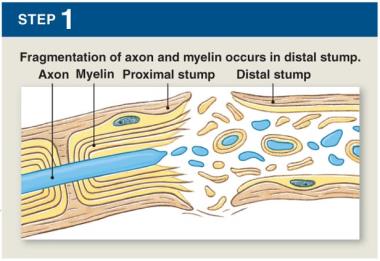
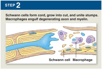
Peripheral nerve regeneration follows a series of steps involving Schwann cells and macrophages:
Fragmentation: Axon and myelin break down in the distal stump.
Schwann Cell Cord Formation: Schwann cells grow into the cut and unite stumps; macrophages clear debris.
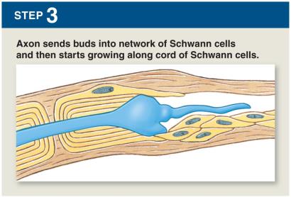
Axonal Budding: Axon sends buds into Schwann cell network and grows along the cord.
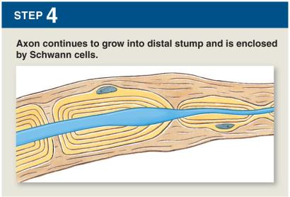
Enclosure: Axon continues to grow into the distal stump and is enclosed by Schwann cells.
Central Nervous System (CNS) Regeneration Limitations
Regeneration in the CNS is limited due to differences in glial cell behavior. Oligodendrocytes, unlike Schwann cells, do not clear myelin debris and may undergo apoptosis or enter a resting state. Astrocytes release chemicals that cause glial scar formation, inhibiting axonal regrowth and re-innervation.
Oligodendrocytes: Do not support regeneration; may form inhibitory scar tissue.
Astrocytes: Release factors that inhibit axonal growth.
Central Chromatolysis
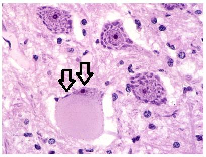
Central chromatolysis is a histopathologic change in the neuron cell body following axonal injury. The nucleus and chromatin are displaced to the cell periphery, and this change may be reversible if regeneration occurs.
Definition: Displacement of nucleus and chromatin in response to axonal injury.
Reversibility: May reverse if axon successfully regenerates.
Stages of Peripheral Nerve Regeneration
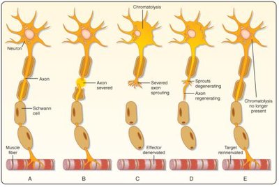
The regeneration process can be visualized in stages, from injury to reinnervation:
Stage A: Healthy neuron and muscle fiber.
Stage B: Axon severed, chromatolysis begins.
Stage C: Effector denervated, axon sprouts.
Stage D: Axon regenerates, target reinnervated.
Stage E: Chromatolysis resolves.
Transmembrane Potential in Neurons
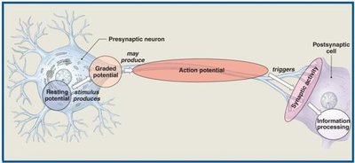
Overview of Neural Activities
Neurons generate electrical signals through ion movements across their plasma membranes. The transmembrane potential is crucial for neural function and is involved in several key processes:
Resting Potential: The baseline electrical charge across the membrane.
Graded Potential: Localized, temporary changes in membrane potential due to stimuli.
Action Potential: Propagated electrical impulse along the axon.
Synaptic Activity: Neurotransmitter release and postsynaptic response.
Information Processing: Integration of stimuli by the postsynaptic cell.
Requirements for Transmembrane Potential
Three main requirements are necessary for establishing and maintaining the transmembrane potential:
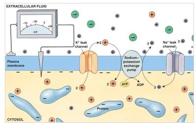
Concentration Gradient: Differences in ion concentrations (Na+, K+, Cl-) across the membrane.
Selective Permeability: Membrane channels allow specific ions to pass.
Charge Difference: Maintenance of electrical potential across the membrane.
Electrochemical Gradient
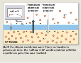
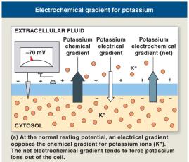
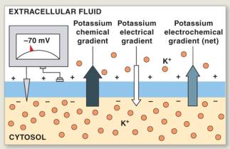
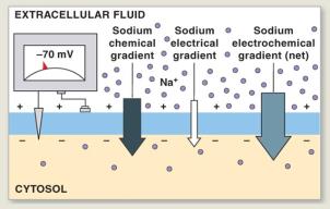
The electrochemical gradient for an ion is the sum of its chemical and electrical gradients across the plasma membrane. This gradient represents potential energy and drives ion movement.
Example: K+ equilibrium potential is –90 mV; Na+ equilibrium potential is +66 mV.
Selective Permeability and Resting Potential
The plasma membrane is highly permeable to K+ ions, so the resting potential of a neuron is close to the equilibrium potential for K+ (–90 mV). However, the actual resting potential is slightly less negative (–70 mV) due to limited Na+ permeability.
Resting Potential: Typically –70 mV in neurons.
K+ Permeability: Major determinant of resting potential.
Na+ Permeability: Small effect due to low permeability.
Membrane Channels: Passive and Active
Membrane permeability to Na+ and K+ is regulated by channels, which can be passive (leak) or active (gated):
Passive Channels: Always open; permeability varies with conditions.
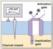
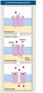
Active (Gated) Channels: Open or close in response to stimuli; most are closed at rest.
Types of Gated Channels
There are three main classes of gated channels:
Chemically Gated Channels: Open in response to specific chemicals (e.g., acetylcholine); found on neuron cell bodies and dendrites.
Voltage Gated Channels: Respond to changes in transmembrane potential; found in axons, skeletal muscle, and cardiac muscle.
Mechanically Gated Channels: Open in response to membrane distortion; found in sensory receptors for touch, pressure, and vibration.
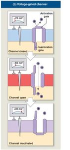
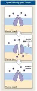
Transmembrane Potential Changes
Resting State and Graded Potentials
At rest, the neuron maintains its resting potential. When exposed to a stimulus, Na+ channels open, producing a graded potential—a localized change in membrane potential that cannot spread far from the site of stimulation.
Graded Potentials: Local changes; magnitude depends on stimulus strength.
Depolarization: Shift toward 0 mV due to Na+ influx.
Depolarization, Repolarization, and Hyperpolarization
These are key changes in membrane potential:
Depolarization: Na+ enters cell, membrane potential becomes less negative.
Repolarization: K+ channels open, membrane potential returns to normal.
Hyperpolarization: K+ leaves cell, membrane potential becomes more negative than resting.
Characteristics of Graded Potentials
Graded potentials share several characteristics:
Local Effect: Most pronounced at site of stimulation; effect decreases with distance.
Passive Spread: Effect spreads passively via local currents.
Depolarization or Hyperpolarization: Depends on channel properties.
Stimulus Strength: Stronger stimulus produces greater change and larger affected area.
Summary Table: Comparison of Channel Types
Channel Type | Stimulus | Location |
|---|---|---|
Chemically Gated | Specific chemicals (e.g., ACh) | Neuron cell body, dendrites |
Voltage Gated | Change in membrane potential | Axons, skeletal muscle, cardiac muscle |
Mechanically Gated | Membrane distortion | Sensory receptors |
Key Equations
Equilibrium potential for an ion (Nernst equation):
Resting membrane potential (Goldman equation):
Additional info: Academic context was added to clarify the steps of nerve regeneration, the role of Schwann cells, and the mechanisms of transmembrane potential changes. Equations were provided for equilibrium and resting membrane potentials.
