 Indietro
Peripheral Nervous System: Anatomy, Plexuses, and Functional Organization
Guida di studio - Note intelligenti
 Appunti personalizzati basati sui tuoi materiali, ampliati con definizioni chiave, esempi e contesto.
Peripheral Nervous System (PNS)
Overview of the Peripheral Nervous System
The Peripheral Nervous System (PNS) consists of nerves and ganglia outside the central nervous system (CNS). It connects the CNS to limbs and organs, facilitating sensory input and motor output.
Nerves: Bundles of axons that transmit signals between the CNS and the rest of the body.
Ganglia: Collections of neuron cell bodies located outside the CNS.
Mixed nerves: Most spinal nerves and some cranial nerves carry both sensory and motor fibers.
Anatomy of a Nerve
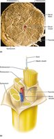
Nerves are complex structures composed of multiple layers of connective tissue and bundles of axons.
Axons: The long, slender projections of neurons that transmit electrical impulses.
Connective tissue layers:
Endoneurium: Surrounds individual axons.
Perineurium: Surrounds bundles of axons (fascicles).
Epineurium: Surrounds the entire nerve.
Myelinated and nonmyelinated axons: Both types are present in nerves, affecting the speed of signal transmission.
Spinal Nerves: Structure and Function
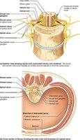
Spinal nerves are mixed nerves that emerge from the spinal cord and branch into rami.
Dorsal root: Contains sensory (afferent) fibers.
Ventral root: Contains motor (efferent) fibers.
Spinal nerve: Formed by the union of dorsal and ventral roots; branches into dorsal and ventral rami.
Dorsal rami: Innervate deep back muscles and skin.
Ventral rami: Innervate the body wall and limbs.
Rami communicantes: Contain autonomic fibers and connect to the sympathetic trunk.
Orientation and Exit of Spinal Nerves
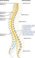
The orientation of spinal nerve roots changes as they descend the spinal cord.
Intervertebral foramen: Spinal nerves exit the vertebral column through these openings.
Cervical spinal nerves: Exit above their corresponding vertebrae.
Lower spinal nerves: Exit below their corresponding vertebrae.
Plexuses of the Peripheral Nervous System
Plexuses are networks of nerves formed by the ventral rami of spinal nerves. They provide redundancy and ensure innervation of limbs and organs.
Cervical plexus: C1-C5; includes the phrenic nerve (C3-C5), which innervates the diaphragm.
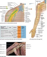
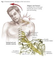
Brachial plexus: C5-T1; supplies the upper limb.
Lumbar plexus: L1-L5; supplies the anterior and medial thigh, leg, and foot.
Sacral plexus: L4-S4; supplies the gluteal region, posterior thigh, leg, and foot.
Brachial Plexus: Structure and Major Nerves
The brachial plexus is a complex network responsible for motor and sensory innervation of the upper limb.
Roots: C5-T1
Trunks: Upper, middle, lower
Cords: Lateral, medial, posterior
Major branches: Musculocutaneous, median, ulnar, axillary, radial nerves
Lumbar Plexus: Structure and Major Nerves
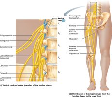
The lumbar plexus provides motor and sensory innervation to the anterior and medial thigh, leg, and foot.
Obturator nerve: L2-L4; medial thigh
Femoral nerve: L2-L4; anterior thigh, medial leg and foot
Lateral femoral cutaneous nerve: L2-L3; lateral thigh
Iliohypogastric and ilioinguinal nerves: L1; anterior pubic/abdominal region
Sacral Plexus: Structure and Major Nerves
The sacral plexus innervates the gluteal region, posterior thigh, leg, and foot.
Sciatic nerve: L4-S3; largest nerve, divides into tibial and common fibular nerves
Tibial nerve: Motor and sensory to posterior leg and plantar foot
Common fibular nerve: Motor and sensory to anterior and lateral leg, dorsal foot
Pudendal nerve: S2-S4; perineum and external anal sphincter
Superior and inferior gluteal nerves: Motor to gluteal muscles
Intercostal Nerves
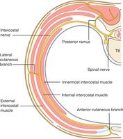
The anterior rami between the brachial and lumbar plexuses form intercostal nerves, which supply the thoracic and abdominal walls.
Segmental supply: Each intercostal nerve innervates a specific region of the thoracic wall.
Muscular and Cutaneous Nerves
Spinal nerves branch into muscular and cutaneous nerves.
Muscular branches: Innervate muscles.
Cutaneous branches: Innervate skin; are superficial to muscular branches.
Dermatomes
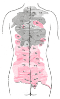
Dermatomes are regions of skin innervated by a single spinal nerve. They are important for clinical diagnosis of nerve injuries.
Dermatome map: Shows the distribution of spinal nerves across the skin.
Difference from cutaneous nerve distribution: Dermatomes are based on spinal nerve origin, while cutaneous nerves may combine fibers from multiple spinal nerves.
Summary Table: Major Plexuses and Their Nerves
Plexus | Major Nerves | Regions Innervated |
|---|---|---|
Cervical | Phrenic | Diaphragm |
Brachial | Musculocutaneous, Median, Ulnar, Axillary, Radial | Upper limb |
Lumbar | Femoral, Obturator, Lateral femoral cutaneous | Anterior/medial thigh, leg, foot |
Sacral | Sciatic, Tibial, Common fibular, Pudendal | Gluteal region, posterior thigh, leg, foot |
Clinical Relevance: Brachial Plexus Injuries
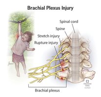
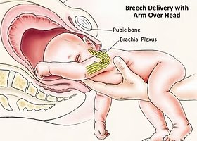
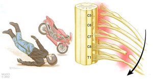
Injuries to the brachial plexus can result in weakness or paralysis of the upper limb.
Causes: Stretching, trauma, or compression (e.g., football tackle, cycling accident, birth injury).
Symptoms: Loss of motor and sensory function in the upper limb.
Key Terms and Definitions
Somatic afferent: Sensory fibers from skin, muscles, and joints.
Somatic efferent: Motor fibers to skeletal muscles.
Visceral afferent: Sensory fibers from internal organs.
Visceral efferent: Motor fibers to smooth muscle, cardiac muscle, and glands.
Fascicle: Bundle of axons within a nerve.
Dermatome: Area of skin supplied by a single spinal nerve.
Additional info:
Redundancy in plexuses ensures that damage to a single spinal nerve does not result in complete loss of function to a region.
Clinical testing of dermatomes helps localize spinal nerve injuries.
