 Indietro
Peripheral Nervous System: Sensory Receptors and Reflex Arcs
Guida di studio - Note intelligenti
 Appunti personalizzati basati sui tuoi materiali, ampliati con definizioni chiave, esempi e contesto.
Peripheral Nervous System: Sensory Receptors and Reflex Arcs
Overview of Sensory Receptors
The peripheral nervous system (PNS) is responsible for transmitting sensory information from the body to the central nervous system (CNS). Sensory receptors are specialized cells or nerve endings that detect changes in the environment and initiate neural signals. These receptors are classified by the type of stimulus they detect, their location in the body, and their structural complexity.
Classification by Stimulus Type: Mechanoreceptors, thermoreceptors, photoreceptors, chemoreceptors, and nociceptors.
Classification by Location: Exteroceptors (external stimuli), interoceptors (internal stimuli), and proprioceptors (body position).
Classification by Structure: Free nerve endings (unencapsulated) and encapsulated nerve endings.
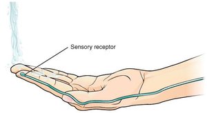
Types of Sensory Receptors
Sensory receptors are specialized to detect specific types of stimuli:
Mechanoreceptors: Detect touch, pressure, vibration, and stretch.
Thermoreceptors: Detect temperature changes.
Photoreceptors: Detect light (primarily in the eyes).
Chemoreceptors: Detect chemical changes in solution (e.g., pH, CO2 levels).
Nociceptors: Detect potentially damaging stimuli that result in pain.
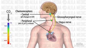
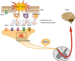
Structural Classification of Sensory Receptors
Sensory receptors differ mainly by whether their nerve endings are bare or wrapped in a protective connective tissue capsule. This structural difference affects their function and adaptation properties.
Free Nerve Endings: Unspecialized, unencapsulated sensory nerve terminals that detect pain, temperature, mechanical pressure, and touch. A-delta fibers are fast-adapting (phasic), while C fibers are slow-adapting (tonic).
Encapsulated Receptors: Include Merkel discs, Meissner's corpuscles, Pacinian corpuscles, and Ruffini endings, each specialized for different types of touch and proprioception.
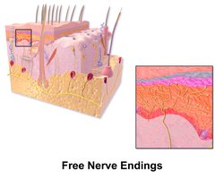
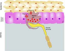
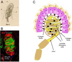
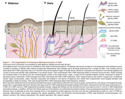
Functional Classification and Location of Sensory Receptors
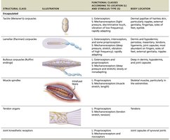
The functional classes of sensory receptors are summarized in the following table, which includes their structural class, illustration, function, and body location:
Structural Class | Illustration | Functional Classes & Stimulus Type | Body Location |
|---|---|---|---|
Tactile (Meissner's) corpuscle | Encapsulated | Mechanoreceptors (light touch, vibration) | Dermal papillae of fingertips, palms, soles |
Lamellar (Pacinian) corpuscle | Encapsulated | Mechanoreceptors (deep pressure, vibration) | Dermis, hypodermis, periosteum, mesentery |
Ruffini corpuscle | Encapsulated | Mechanoreceptors (stretch) | Deep dermis, joint capsules, ligaments |
Muscle spindle | Encapsulated | Proprioceptors (muscle stretch) | Skeletal muscles |
Tendon organ | Encapsulated | Proprioceptors (tendon tension) | Tendons |
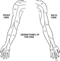
Dermatomes and Sensory Innervation
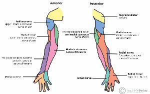
Dermatomes are regions of skin innervated by specific spinal nerves. Understanding dermatomes is essential for diagnosing nerve injuries and sensory deficits.
Dermatomes of the Arm: Each area of the arm is supplied by a specific spinal nerve root (e.g., C5, C6, C7, C8, T1).
Clinical Application: Sensory complaints (e.g., thumb, shoulder, pinky pain) can be mapped to specific dermatomes for differential diagnosis.
Somatic Reflex Arcs
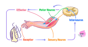
Reflex arcs are neural pathways that mediate automatic responses to stimuli, helping maintain homeostasis. They consist of sensory receptors, sensory neurons, interneurons, motor neurons, and effectors.
Components: Receptor, sensory neuron, interneuron, motor neuron, effector.
Actions: Agonist muscles contract, antagonist muscles relax (or vice versa).
Function: Reflexes protect the body and regulate muscle tone.
Proprioceptors: Muscle Spindle and Golgi Tendon Organ
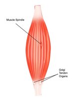
Proprioceptors are specialized sensory receptors that provide information about body position and movement. The muscle spindle senses muscle stretch and speed, while the Golgi tendon organ senses tendon tension.
Muscle Spindle: Located within skeletal muscles; detects changes in muscle length and speed of stretch.
Golgi Tendon Organ: Located in tendons; detects tension and prevents excessive contraction.
Reflexes: Muscle Stretch and Tendon Reflex
Muscle stretch reflexes and tendon reflexes are essential for maintaining muscle tone and preventing injury.
Muscle Stretch Reflex: Tapping a ligament stretches the muscle, activating the muscle spindle. The afferent signal triggers contraction of the agonist muscle and inhibition of the antagonist muscle via interneurons.
Tendon Reflex: Muscle contraction activates the Golgi tendon organ, which sends afferent signals to interneurons. These interneurons excite the antagonist muscle and inhibit the contracting muscle, preventing excessive tension.
Sensory Adaptation
Sensory adaptation is the process by which sensory receptors become less responsive to a constant stimulus over time. This is important for filtering out non-threatening stimuli and focusing on changes in the environment.
Phasic Receptors: Fast-adapting; signal changes in the environment (e.g., Meissner's corpuscles).
Tonic Receptors: Slow-adapting or nonadapting; provide sustained responses for continuous, protective information (e.g., pain, body position).
Example: The sensation of an itchy sweater fades over time due to adaptation by phasic receptors.
Repair of PNS Neurons
Peripheral nervous system neurons can repair after injury, a process guided by Schwann cells. The steps include axon degeneration, cleanup of damaged tissue, axon regrowth, and remyelination.
Schwann Cells: Clear damaged tissue, guide axon regrowth, and restore the myelin sheath.
Key Takeaway: PNS neurons can regenerate, but CNS neurons cannot.
Summary Table: Sensory Receptor Types
Receptor Type | Stimulus Detected | Location | Adaptation |
|---|---|---|---|
Free Nerve Endings | Pain, temperature, touch | Skin, mucous membranes | Variable (phasic or tonic) |
Merkel Discs | Light touch | Basal epidermis, hair follicles | Slowly adapting |
Meissner's Corpuscles | Light touch, vibration | Dermal papillae | Rapidly adapting |
Pacinian Corpuscles | Deep pressure, vibration | Deep dermis, hypodermis | Rapidly adapting |
Ruffini Endings | Stretch | Deep dermis, ligaments | Slowly adapting |
Muscle Spindle | Muscle stretch | Skeletal muscle | Slowly adapting |
Golgi Tendon Organ | Tendon tension | Tendons | Slowly adapting |
Key Concepts and Takeaways
Receptors are classified by function, location, and structure.
Adaptation is more likely in benign senses (light touch, vibration) than in protective senses (pain, body position).
Reflexes are spinal cord-mediated reactions that help maintain homeostasis.
CNS neurons cannot repair; PNS neurons repair slowly.
