 Indietro
Peripheral Nervous System: Sensory Receptors, Pain, Nerve Repair, Cranial Nerves, and Reflexes
Guida di studio - Note intelligenti
 Appunti personalizzati basati sui tuoi materiali, ampliati con definizioni chiave, esempi e contesto.
Peripheral Nervous System
Overview
The peripheral nervous system (PNS) connects the central nervous system (CNS) to limbs and organs, serving as a communication relay. This section covers the organization of the PNS, sensory receptors, pain mechanisms, nerve repair, cranial nerves, and somatic reflexes.
Organization of the Nervous System
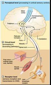
Levels of Sensory Processing
Sensory information is processed through three hierarchical levels:
Receptor Level: Sensory reception and transmission to the CNS. Examples include joint kinesthetic receptors, muscle spindles, and free nerve endings for pain, cold, and warmth.
Circuit Level: Processing in ascending pathways from receptors through the spinal cord, cerebellum, or reticular formation to the thalamus.
Perceptual Level: Processing in cortical sensory centers, where pathways from the thalamus lead to the motor cortex or somatosensory cortex.
Sensory Receptors
Classification by Stimulus Type
Sensory receptors are specialized to detect specific types of stimuli:
Mechanoreceptors: Respond to mechanical force (touch, pressure, vibration, stretch).
Thermoreceptors: Respond to temperature changes.
Photoreceptors: Respond to light (e.g., retina of the eye).
Chemoreceptors: Respond to chemicals in solution (smell, taste, changes in blood/interstitial fluid chemistry).
Nociceptors: Respond to damaging stimuli that result in pain.
Classification by Location
Exteroceptors: Detect external stimuli (touch, pressure, pain, temperature).
Interoceptors: Detect internal stimuli (chemical changes, tissue stretch).
Proprioceptors: Detect internal stimuli in skeletal muscles, tendons, joints, and ligaments, providing information about body position.
Receptors and Sensations
Sensory receptor: Specialized to detect stimuli, ranging from bare nerve endings to complex sense organs.
Sense organ: Combines nervous, muscular, epithelial, or connective tissue to enhance response to stimuli.
Sensation: Occurs when a receptor is excited by a stimulus and sensory signals are sent to the brain for processing.
Awareness of sensation depends on whether the brain processes the signal consciously.
Types of Sensory Information
Type (Modality)
The modality refers to the kind of environmental stimulus (e.g., vision, taste, pain, touch). The brain distinguishes modalities based on where sensory signals terminate in the brain.
Location
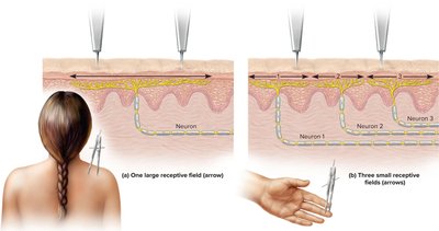
The location of a stimulus is determined by which sensory neuron sends a signal to the brain. Each neuron has a receptive field—an area where stimuli can activate it. The brain cannot distinguish the exact point within a receptive field that was stimulated.
Intensity
Intensity is determined by:
Which nerves are sending signals (sensitive or not)
How many nerves are sending signals
The frequency of nerve signals
Duration
Duration refers to how long a stimulus lasts. All receptors can adapt to prolonged stimuli, decreasing their firing rate over time (sensory adaptation). Some adapt quickly (e.g., smell), while others adapt slowly (e.g., pain).
Sensory Transduction
Sensory organs act as transducers, converting one form of energy (light, heat, chemicals, touch, sound) into another (action potentials). This process is called sensory transduction.
Pain
Mechanisms of Pain
Pain: Discomfort caused by tissue damage or noxious stimulation; can be visceral or somatic.
Nociceptors: Specialized nerve endings that respond to chemicals released by injured tissues; concentrated in skin, mucous membranes, and most organs (not present in the brain).
Types of Pain
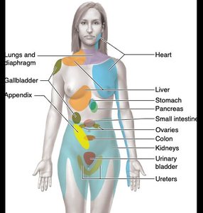
Referred Pain: Visceral pain perceived as originating from the skin (e.g., left arm pain during a heart attack).
Phantom Pain: Sensation of pain from an amputated limb, likely due to missing sensory input in the brain and spinal cord.
Spinal Gating: Neurons in the spinal cord and brainstem secrete endorphins and enkephalins, blocking pain signals before they reach consciousness.


Nerve Repair
Regeneration in the CNS vs. PNS
CNS Nerves: Cannot regenerate due to inhibitory factors from oligodendrocytes and scar tissue formation by astrocytes.
PNS Nerves: Can regenerate with the help of Schwann cells, which clean up debris, release growth factors, and form new myelin sheaths.
Cranial Nerves
Overview of the 12 Cranial Nerves
Cranial nerves are numbered I–XII and have sensory, motor, or mixed functions:
Number | Name | Function |
|---|---|---|
I | Olfactory | Purely sensory; smell |
II | Optic | Purely sensory; vision |
III | Oculomotor | Mainly motor; eye movement |
IV | Trochlear | Primarily motor; eye muscles |
V | Trigeminal | Main sensory nerve of the face |
VI | Abducens | Primarily motor; eye muscles |
VII | Facial | Chief motor nerve of the face |
VIII | Vestibulocochlear | Mostly sensory; balance and hearing |
IX | Glossopharyngeal | Mixed; tongue and pharynx |
X | Vagus | Mixed; parasympathetic regulation of heart, breathing, digestion |
XI | Accessory | Mixed; primarily motor to trapezius and sternocleidomastoid |
XII | Hypoglossal | Mixed; primarily motor for tongue and swallowing |
Somatic Reflexes
Reflex Arc
A reflex is a quick, involuntary reaction of a gland or muscle to a stimulus. Reflexes have four key properties:
Quick: Simple neural pathways with few interneurons.
Involuntary: Occur without conscious awareness and cannot be suppressed.
Stereotyped: Occur the same way every time.
Stimulation: Always a response to sensory input.
Types of Reflexes
Visceral Reflexes: Involve cardiac muscle, smooth muscle, or glands (e.g., increased heart rate in fear, constriction of esophagus when swallowing).
Somatic Reflexes: Involve skeletal muscle (e.g., knee-jerk reflex, pulling hand away from a hot stove).
Somatic Reflex Arc Pathway
Receptor: Sensory nerve ending or sense organ in skin, muscle, or tendon.
Afferent (sensory) neuron: Carries signal to spinal cord or brainstem.
Integrating center: Synaptic contact in gray matter; may involve interneurons.
Efferent (motor) neuron: Carries motor signal to skeletal muscle.
Effector: Skeletal muscle or organ that carries out the response.
Examples of Somatic Reflexes
Stretch Reflex (Knee-Jerk): Doctor taps patellar tendon; muscle spindles detect stretch, signal travels via afferent neuron to spinal cord, synapses with motor neuron, causing quadriceps contraction (monosynaptic reflex arc).
Flexor (Withdrawal) Reflex: Protective reflex involving multiple muscles; polysynaptic (involves interneurons). For example, withdrawing foot from pain excites hamstring and inhibits quadriceps.
Babinski’s Sign
If the primary motor cortex or corticospinal tract is damaged, the plantar reflex is replaced by Babinski’s sign (great toe dorsiflexes, smaller toes fan laterally). Infants normally exhibit this reflex until about 1 year of age.
Sample Question
The main sensory nerve of the face is the?
A. abducens
B. accessory
C. trigeminal
D. facial
