Indietro
Peripheral Nervous System: Structure, Function, and Cranial Nerves
Guida di studio - Note intelligenti
 Appunti personalizzati basati sui tuoi materiali, ampliati con definizioni chiave, esempi e contesto.
Peripheral Nervous System (PNS)
Overview and Functional Divisions
The Peripheral Nervous System (PNS) connects the Central Nervous System (CNS) to the rest of the body and the external environment. It is functionally divided into sensory and motor divisions, each with somatic and visceral components.
Sensory Division: Transmits sensory information from the body to the CNS.
Somatic Sensory Division: Carries information from skin, muscles, and joints.
Visceral Sensory Division: Carries information from internal organs.
Motor Division: Transmits motor commands from the CNS to effectors.
Somatic Motor Division: Controls voluntary movements of skeletal muscles.
Visceral Motor Division (Autonomic Nervous System, ANS): Controls involuntary functions; includes the Sympathetic (fight or flight) and Parasympathetic (rest and digest) systems.
Peripheral Nerves
Structure and Classification
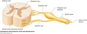
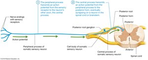
Peripheral nerves are classified as mixed, sensory, or motor nerves. Spinal nerves (31 pairs) and cranial nerves (12 pairs) connect the PNS to the CNS. Each spinal nerve is formed by the union of anterior and posterior roots, with the posterior root ganglion housing sensory neuron cell bodies.
Mixed Nerves: Contain both sensory and motor fibers.
Sensory Nerves: Carry sensory information only.
Motor Nerves: Carry motor commands only.
Nerve Anatomy
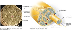
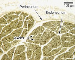
Nerves are organized into bundles called fascicles, each surrounded by connective tissue layers:
Epineurium: Outermost layer, surrounds the entire nerve.
Perineurium: Surrounds each fascicle.
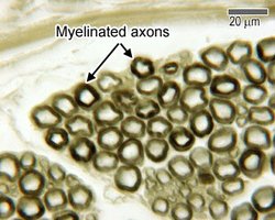
Endoneurium: Surrounds individual axons within a fascicle.
Functional Overview of the PNS
Sensory and Motor Integration
The PNS works closely with the CNS to detect stimuli and initiate appropriate responses. Sensory neurons transmit information to the cerebral cortex, where it is processed and interpreted. Motor commands are then sent from the cortex to lower motor neurons, which stimulate muscle contractions.
Cranial Nerves
Classification and Functions
Cranial nerves are classified as sensory, motor, or mixed, and each has specific functions related to sensation, movement, or both. There are 12 pairs, each with unique roles in the head, neck, and some visceral organs.
Mnemonic for Cranial Nerves: "Oh Once One Takes The Anatomy Final Very Good Vacations Are Happening" (I-XII).
Mnemonic for Function: "Some Say Money Matters But My Brother Says Big Brains Matter More" (Sensory, Motor, Both).
Motor Cranial Nerves
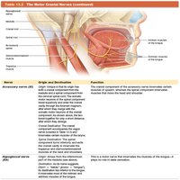
Motor cranial nerves control movements of muscles in the head, neck, and tongue.
Mixed Cranial Nerves
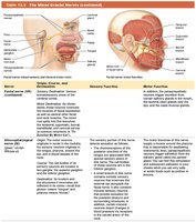
Mixed cranial nerves have both sensory and motor functions, such as the facial and glossopharyngeal nerves.
Cranial Nerve Distribution
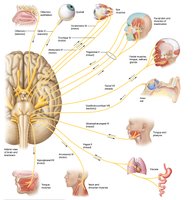
The cranial nerves innervate various structures, including the eyes, face, tongue, and internal organs.
Spinal Nerves and Plexuses
Formation and Classification
There are 31 pairs of spinal nerves, classified by region: cervical, thoracic, lumbar, sacral, and coccygeal. Most ventral rami form plexuses, which are networks of nerves that supply specific regions of the body.
Cervical Plexus: C1-C5
Brachial Plexus: C5-T1
Lumbar Plexus: L1-L4
Sacral Plexus: L4-S4
Coccygeal Plexus: S4-S5
Sensory Transduction and Receptors
Role of PNS in Sensation
Sensory transduction is the process by which sensory receptors convert stimuli into electrical signals. Mechanically gated ion channels open in response to pressure, allowing Na+ to enter and generate a receptor potential.
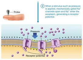
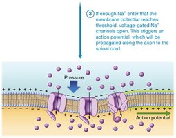
Types of Sensory Receptors
Sensory receptors are classified by structure, location, and stimulus type:
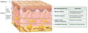
Encapsulated Nerve Endings: Surrounded by connective tissue (e.g., Meissner corpuscles).
Free Nerve Endings: Not surrounded by connective tissue (e.g., hair follicle receptors).
Exteroceptors: Detect external stimuli.
Interoceptors: Detect internal stimuli.
Proprioceptors: Monitor body position.
Thermoreceptors: Detect temperature changes.
Chemoreceptors: Detect chemical changes.
Photoreceptors: Detect light.
Nociceptors: Detect pain.
Mechanoreceptors: Detect mechanical changes; examples include Merkel cells, Meissner corpuscles, Ruffini endings, and Pacinian corpuscles.
Somatic Sensory Neurons
Structure and Function
First-order somatic sensory neurons are pseudounipolar, with a cell body, peripheral process, and central process. They transmit sensory information from receptors to the CNS.
Transduction Factors and Receptive Fields
Speed of Conduction and Receptive Fields
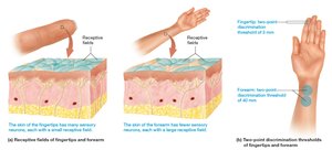
The speed of action potential conduction depends on axon diameter and myelin thickness. The receptive field is the area served by a single neuron; smaller fields allow for finer discrimination.
Dermatomes and Referred Pain
Dermatome Maps and Clinical Relevance
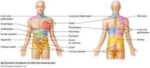
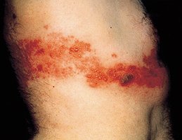
Dermatomes are skin segments supplied by specific spinal nerves. Dermatome maps are used clinically to assess sensory pathway integrity. Referred pain is often felt along the dermatome of the affected nerve.
Reflex Arcs and Types of Reflexes
Classification and Mechanisms
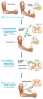
Reflexes are classified by the number of synapses (monosynaptic or polysynaptic) and the organ involved (somatic or visceral). Types include simple stretch, Golgi tendon, flexion (withdrawal), crossed-extension, and cranial nerve reflexes.
Stretch Receptors in Skeletal Muscles
Muscle Spindles and Golgi Tendon Organs
Mechanoreceptors in muscles and tendons monitor muscle length and force. Muscle spindles detect stretch, while Golgi tendon organs detect tension at the muscle-tendon junction.
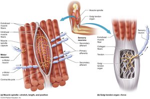
Stretch and Golgi Tendon Reflexes
Mechanisms and Functions
Stretch reflexes maintain muscle tone and posture, while Golgi tendon reflexes protect muscles from excessive tension by causing relaxation.
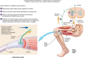
Withdrawal and Crossed Extensor Reflexes
Protective Reflexes
The flexion (withdrawal) reflex removes a body part from harmful stimuli, while the crossed extensor reflex maintains balance by activating opposite limb muscles.
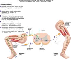
Cranial Nerve Details
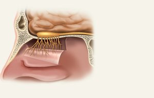
Olfactory Nerve (I)
Responsible for the sense of smell. Damage impairs olfaction.
Optic Nerve (II)
Provides vision. Damage causes blindness in part or all of the visual field.
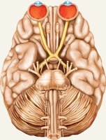
Oculomotor Nerve (III)
Controls eye movements, iris, lens, and upper eyelid. Damage causes drooping eyelid, dilated pupil, double vision, and difficulty focusing.
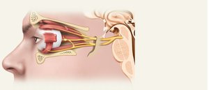
Trochlear Nerve (IV)
Controls superior oblique muscle for eye movement. Damage causes double vision and inability to rotate eye inferolaterally.
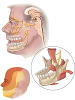
Trigeminal Nerve (V)
Largest cranial nerve; major sensory nerve of the face. Has three divisions: ophthalmic (V1), maxillary (V2), and mandibular (V3).
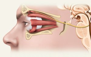
Abducens Nerve (VI)
Controls lateral rectus muscle for eye movement. Damage results in inability to rotate eye laterally.
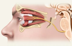
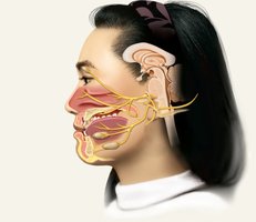
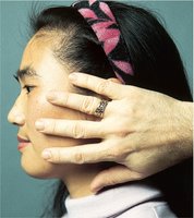
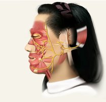
Facial Nerve (VII)
Major motor nerve of facial muscles; also involved in taste and glandular secretion. Damage produces sagging facial muscles and disturbed taste.
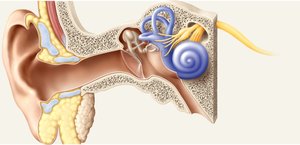
Vestibulocochlear Nerve (VIII)
Nerve of hearing and equilibrium. Damage produces deafness, dizziness, and loss of balance.
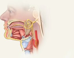
Glossopharyngeal Nerve (IX)
Involved in swallowing, salivation, taste, and regulation of blood pressure and respiration. Damage results in loss of taste and impaired swallowing.
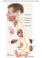
Vagus Nerve (X)
Most extensive distribution; controls cardiac, pulmonary, digestive, and urinary functions. Damage can be fatal if both nerves are cut.
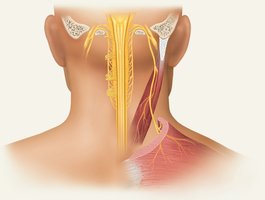
Accessory Nerve (XI)
Controls swallowing, head, neck, and shoulder movement. Damage impairs these movements.
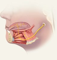
Hypoglossal Nerve (XII)
Controls tongue movements for speech, food manipulation, and swallowing. Damage causes tongue deviation and atrophy.