 Indietro
Chap 13: Peripheral Nervous System - McNeese, Duck
Guida di studio - Note intelligenti
 Appunti personalizzati basati sui tuoi materiali, ampliati con definizioni chiave, esempi e contesto.
The Peripheral Nervous System (PNS)
Overview and Structural Organization
The Peripheral Nervous System (PNS) serves as the communication link between the central nervous system (CNS) and the external/internal environment. It consists of all neural structures outside the brain and spinal cord, and is organized into sensory and motor divisions, each with further subdivisions.
Sensory (afferent) division: Transmits sensory information from receptors to the CNS.
Motor (efferent) division: Carries motor commands from the CNS to effectors (muscles and glands).
Somatic nervous system: Controls voluntary movements.
Autonomic nervous system (ANS): Regulates involuntary functions, subdivided into sympathetic and parasympathetic divisions.
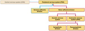
Components of the PNS
Sensory Receptors and Sensations
Nerves and Their Structure and Repair
Motor Endings and Motor Activity
Reflex Activity
Sensory Receptors
Classification and Function
Sensory receptors are specialized to respond to environmental changes (stimuli). Their activation results in graded potentials that trigger nerve impulses, leading to sensation (awareness) and perception (interpretation) in the brain.
Mechanoreceptors: Respond to touch, pressure, vibration, and stretch.
Thermoreceptors: Sensitive to temperature changes.
Photoreceptors: Respond to light energy (e.g., retina).
Chemoreceptors: Respond to chemicals (e.g., smell, taste, blood chemistry).
Nociceptors: Sensitive to pain-causing stimuli (e.g., extreme heat/cold, pressure, chemicals).
Classification by Structure
Sensory receptors are categorized as simple (general senses) or complex (special senses). Simple receptors are modified dendritic endings of sensory neurons and are further divided into nonencapsulated (free) and encapsulated types.
Nonencapsulated (free) nerve endings: Found in epithelia and connective tissues; detect tactile sensations, temperature, pain, and muscle sense.
Encapsulated nerve endings: Dendritic endings encased in connective tissue; mostly mechanoreceptors.
Special sense receptors: Located in complex sense organs for vision, hearing, equilibrium, smell, and taste.
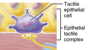
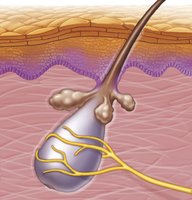
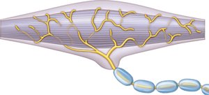
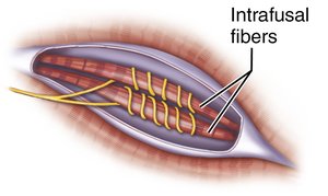
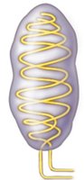
Sensory Processing
Levels of Neural Integration
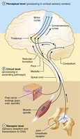
The somatosensory system processes sensory information from the body wall and limbs. It involves three levels of neural integration:
Receptor Level: Sensory receptors generate graded potentials in response to stimuli, which may lead to action potentials.
Phasic receptors: Fast-adapting; signal at the beginning or end of a stimulus (e.g., touch, pressure).
Tonic receptors: Slow-adapting or non-adapting; maintain response (e.g., pain, proprioception).
Circuit Level: Involves first, second, and third order neurons transmitting signals to the cortex.
Perceptual Level: Involves summation, frequency, pattern recognition, and identification of submodalities (e.g., sweet taste).
Perception of Pain
Visceral and Referred Pain
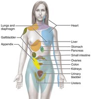
Visceral pain arises from stimulation of visceral organ receptors and is often felt as vague aching, gnawing, or burning. Referred pain occurs when pain from one region is perceived as coming from another, due to shared nerve pathways between visceral and somatic fibers.
Causes: Tissue stretching, ischemia, chemicals, muscle spasms.
Clinical relevance: Understanding referred pain is crucial for diagnosis (e.g., heart attack pain felt in the arm).
Nerves and Ganglia
Structure and Classification of Nerves
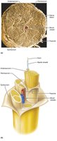
Nerves are bundles of myelinated and nonmyelinated peripheral axons enclosed by connective tissue. They are classified as spinal or cranial, and further as sensory, motor, or mixed nerves.
Sensory (afferent) nerves: Carry information from muscles/organs to the brain.
Motor (efferent) nerves: Transmit commands from the brain to muscles/organs.
Mixed nerves: Contain both sensory and motor fibers; most nerves are mixed.
Ganglia
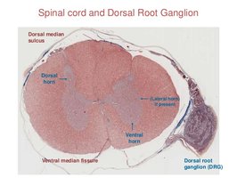
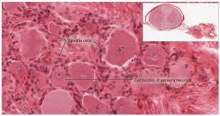
Ganglia are clusters of neuron cell bodies in the PNS. Afferent ganglia (dorsal root ganglia) contain sensory neuron cell bodies, while efferent ganglia (autonomic ganglia) contain autonomic motor neuron cell bodies.
Cranial Nerves
Overview and Functions
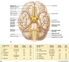
There are 12 pairs of cranial nerves, most of which are mixed, except for two pairs that are purely sensory. They originate from the forebrain or brainstem and serve various sensory and motor functions.
Selected Cranial Nerves
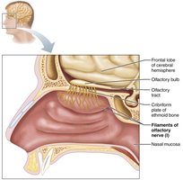
Olfactory (I): Sensory nerves of smell; terminate in the primary olfactory cortex.
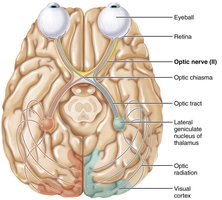
Optic (II): Sensory nerves of vision; pathway includes optic chiasma and visual cortex.
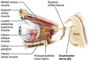
Oculomotor (III): Motor nerves for eye movement, eyelid, iris, and lens shape.
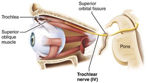
Trochlear (IV): Motor nerve for superior oblique muscle of the eye.
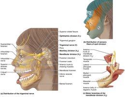
Trigeminal (V): Largest cranial nerve; sensory from face, motor for mastication.
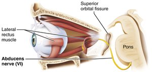
Abducens (VI): Motor nerve for lateral rectus muscle of the eye.
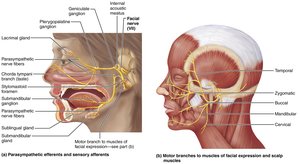
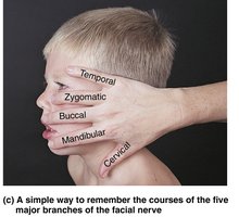
Facial (VII): Motor for facial expression, sensory for taste; five major branches.
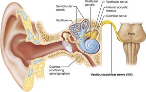
Vestibulocochlear (VIII): Sensory for hearing and equilibrium.
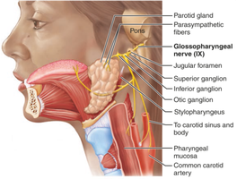
Glossopharyngeal (IX): Motor for swallowing, sensory for taste and carotid receptors.
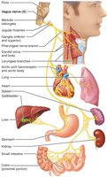
Vagus (X): Only cranial nerve extending beyond head/neck; regulates heart, lungs, abdominal viscera.
Spinal Nerves
Structure and Distribution
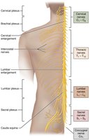
There are 31 pairs of spinal nerves, all mixed, supplying all body parts except the head and part of the neck. They are grouped as cervical, thoracic, lumbar, sacral, and coccygeal.
8 pairs cervical
12 pairs thoracic
5 pairs lumbar
5 pairs sacral
1 pair coccygeal
Formation and Branching of Spinal Nerves
Each spinal nerve connects to the spinal cord via ventral (motor) and dorsal (sensory) roots. After exiting the vertebral foramen, spinal nerves divide into dorsal ramus, ventral ramus, meningeal branch, and rami communicantes (autonomic fibers).
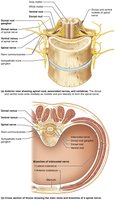
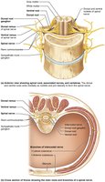
Nerve Plexuses
Innervation of Specific Body Regions
Nerve plexuses are interlacing networks formed by ventral rami in cervical, brachial, lumbar, and sacral regions. Plexuses allow each limb muscle to be innervated by more than one spinal nerve, providing redundancy and protection against paralysis.
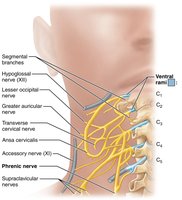
Cervical plexus (C1-C4): Innervates skin and muscles of neck, ear, back of head, and shoulders; includes phrenic nerve.
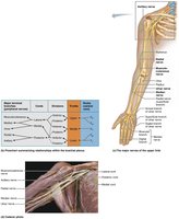
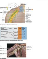
Brachial plexus (C5-T1): Innervates upper limb; includes axillary, musculocutaneous, median, ulnar, and radial nerves.
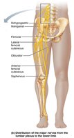
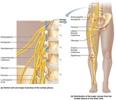
Lumbar plexus (L1-L4): Innervates thigh, abdominal wall, psoas muscle; includes femoral and obturator nerves.
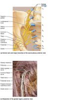
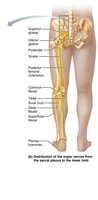
Sacral plexus (L4-S4): Serves buttock, lower limb, pelvic structures, perineum; includes sciatic nerve (tibial and common fibular components).
Anterolateral Thorax and Abdominal Wall
Ventral rami of T1-T12 do not form a plexus; intercostal nerves supply muscles of ribs, thorax, and abdominal wall, with cutaneous branches to the skin.
Summary Table: General Sensory Receptors
The following table summarizes the classification of general sensory receptors by structure and function:
Type | Structure | Function |
|---|---|---|
Nonencapsulated | Free nerve endings | Tactile, temperature, pain, muscle sense |
Encapsulated | Connective tissue capsule | Mostly mechanoreceptors |
Special senses | Complex sense organs | Vision, hearing, equilibrium, smell, taste |
Key Equations:
Graded potential to action potential conversion:
Additional info: Academic context was added to clarify the structure and function of the PNS, sensory processing, and nerve plexuses, as well as to provide clinical relevance for pain perception and redundancy in nerve supply.
