 Indietro
Chapter 13 Peripheral Nervous System: Structure, Function, and Clinical Relevance
Guida di studio - Note intelligenti
 Appunti personalizzati basati sui tuoi materiali, ampliati con definizioni chiave, esempi e contesto.
Peripheral Nervous System (PNS) Overview
Introduction to the PNS
The Peripheral Nervous System (PNS) serves as the critical link between the Central Nervous System (CNS) and the rest of the body, facilitating both sensory input and motor output. It detects external and internal stimuli and transmits information to and from the CNS.
Sensory Input: PNS detects sensory stimuli and delivers them to the CNS.
Motor Output: CNS processes input and sends impulses through the PNS to effectors (muscles and glands).
Spinal and Cranial Nerves: Both belong to the PNS, even though they attach directly to the spinal cord and brain.
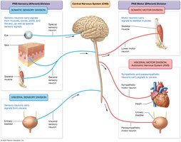
Divisions of the PNS
The PNS is divided into sensory (afferent) and motor (efferent) divisions, each with somatic and visceral components.
Sensory (Afferent) Division:
Somatic Sensory: Signals from muscles, bones, joints, skin, and special senses.
Visceral Sensory: Signals from internal organs (thoracic and abdominopelvic cavities).
Motor (Efferent) Division:
Somatic Motor: Signals to skeletal muscles.
Visceral Motor (Autonomic Nervous System): Signals to cardiac and smooth muscles and glands.
Sympathetic: "Fight or Flight" system.
Parasympathetic: "Rest and Digest" system.
Peripheral Nerves and Associated Ganglia
Structure and Function of Peripheral Nerves
Peripheral nerves are bundles of axons bound by connective tissue sheaths and innervate most body structures.
Mixed Nerves: Contain both sensory and motor axons.
Sensory Nerves: Only sensory axons.
Motor Nerves: Mostly motor axons, with some sensory axons monitoring muscle stretch and tension.
Cranial Nerves: Attach to the brain; innervate head and neck.
Spinal Nerves: Thirty-one pairs branch from the spinal cord; innervate structures below the neck.
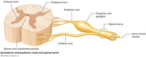
Connective Tissue Sheaths of Spinal Nerves
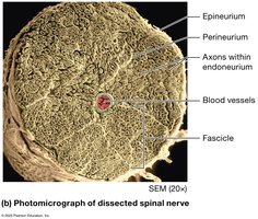
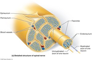
Spinal nerves are organized by three connective tissue layers:
Epineurium: Surrounds each nerve.
Perineurium: Surrounds each fascicle (group of axons).
Endoneurium: Surrounds each individual axon.
Cranial Nerves
Overview and Classification
Cranial nerves are classified as sensory, motor, or mixed, and are essential for sensory and motor functions of the head and neck.
Sensory Only: Olfactory, Optic, Vestibulocochlear
Motor Only: Oculomotor, Trochlear, Abducens, Accessory, Hypoglossal
Mixed: Trigeminal, Facial, Glossopharyngeal, Vagus
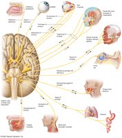
Key Cranial Nerves and Their Functions slide 11-30*
Olfactory (I): Sensory for smell
Optic (II): Sensory for vision
Oculomotor (III), Trochlear (IV), Abducens (VI): Motor for eye movement
Trigeminal (V): Mixed; sensory for face, motor for mastication
Facial (VII): Mixed; sensory for taste, motor for facial expression
Vestibulocochlear (VIII): Sensory for hearing and balance
Glossopharyngeal (IX): Mixed; sensory for taste, motor for swallowing
Vagus (X): Mixed; sensory and motor for viscera
Accessory (XI): Motor for neck muscles
Hypoglossal (XII): Motor for tongue
Spinal Nerves and Plexuses
Structure and Branching of Spinal Nerves
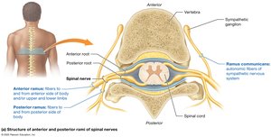
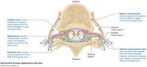
Spinal nerves exit the vertebral column and split into posterior and anterior rami, serving different regions of the body.
Posterior Ramus: travels to the posterior side of the body.
Anterior Ramus: travels to the anterior side of the body and/or upper and lower limbs.
Ramus Communicans: Contain visceral motor orautonomic axons of the sympathetic nervous system; Motoraxons only
31 pairs of Spinal Nerves
8 pairs of Cervical nerves (C1 -C 8)
12 pairs of Thoracic nerves (T1 -T 12)
5 pairs of Lumbar nerves (L1 -L5 )
5 pairs of Sacral nerves1 5 (S1 -S5 )
1 tiny pair of Coccygeal nerves (Co 1)
Spinal Nerve Plexuses
Cervical Plexus: Supplies neck, head, chest, shoulders, and diaphragm (phrenic nerve).
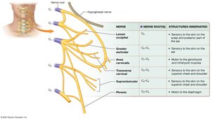
Brachial Plexus: Supplies upper limb; includes axillary, radial, musculocutaneous, median, and ulnar nerves.
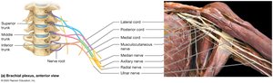
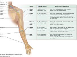
Lumbar Plexus: Supplies pelvis and lower extremity; includes obturator and femoral nerves.
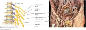
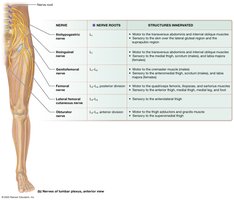
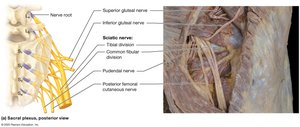
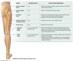
Sacral Plexus: Supplies pelvis, gluteal region, and lower extremity; includes sciatic, tibial, and common fibular nerves.
Sensory Receptors and Sensation
Sensory Transduction and Receptor Types
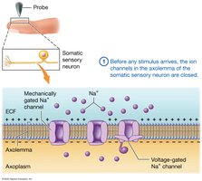
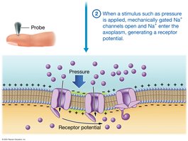
Sensory transduction is the conversion of a stimulus into an electrical signal, beginning at sensory receptors.
Encapsulated Nerve Endings: Surrounded by supporting cells.
Free Nerve Endings: Lack supporting cells.
Classification of Sensory Receptors
By Location:
Exteroceptors: Near body surface; detect external stimuli.
Interoceptors: Within body; detect internal stimuli.
By Stimulus Type:
Mechanoreceptors: Respond to mechanical deformation.
Thermoreceptors: Respond to temperature changes.
Chemoreceptors: Respond to chemicals.
Photoreceptors: Respond to light (eye only).
Nociceptors: Detect pain.
Types of Mechanoreceptors
Tactile (Merkel) Endings: Discriminative touch, form, and texture.
Tactile (Meissner) Corpuscles: Discriminative touch.
Bulbous (Ruffini) Corpuscles: Stretch and movement.
Lamellated (Pacinian) Corpuscles: Vibratory stimuli and deep pressure.
Hair Follicle Receptors: Respond to hair bending.
Proprioceptors: Detect movement and position.
Sensory Neurons and Pathways
Structure and Function of Sensory Neurons
First-order somatic sensory neurons are pseudounipolar, with cell bodies in ganglia and axons that transmit sensory information to the CNS.
Peripheral Process: Long axon with sensory receptors.
Central Process: Travels to CNS.
Receptive Fields and Two-Point Discrimination
Large Receptive Fields: Less sensation, more branching.
Small Receptive Fields: More sensation, less branching.
Two-Point Discrimination: Measures receptive field size.
Dermatomes and Referred Pain
Dermatomes: Skin regions supplied by specific spinal nerves; used clinically to assess sensory pathways.
Referred Pain: Pain from organs perceived as cutaneous pain, pain felt in area that does not necessarily have pain introduced to it
Motor Output and Reflexes
Motor Neurons and Control of Movement
Skeletal muscle contraction is initiated by somatic motor neurons, with upper motor neurons in the CNS relaying signals to lower motor neurons in the PNS.
Alpha (α) Motor Neurons: Stimulate muscle contraction.
Gamma (γ) Motor Neurons: Innervate stretch receptors (intrafusal fibers).
Reflex Arcs and Types of Reflexes
Reflexes are automatic responses to sensory input, often protective.
Monosynaptic Reflex: Single synapse (e.g., stretch reflex).
Polysynaptic Reflex: Multiple synapses.
Somatic Reflex: Involves skeletal muscles.
Visceral Reflex: Involves internal organs.
Stretch Receptors and Golgi Tendon Organs
Muscle Spindles: Detect muscle stretch; more spindles in muscles for fine movement.
Golgi Tendon Organs: Monitor tension; protect muscles and tendons from damage.
Somatic Reflexes
Simple Stretch Reflex: Returns muscle to optimal length (e.g., patellar reflex).
Flexion (Withdrawal) Reflex: Pulls back from painful stimulus.
Crossed-Extension Reflex: Extends opposite limb for balance.
Golgi Tendon Reflex: Relaxes muscle if tension is too high.
Cranial Nerve Reflexes: Gag and corneal blink reflexes.
Clinical Relevance: Sensory and Motor Neuron Disorders
Peripheral Neuropathies
Disorders affecting sensory and motor neurons of the PNS can result in paralysis, weakness, or abnormal reflexes.
Lower Motor Neuron Disorders: Injury to nerves or cell bodies; may cause paralysis or paresis.
Upper Motor Neuron Disorders: Affect CNS pathways; may cause spinal shock, spasticity, and Babinski sign.
Amyotrophic Lateral Sclerosis (ALS)
ALS involves degeneration of motor neurons, leading to muscle weakness and eventual death.
Symptoms: Muscle weakness, upper motor neuron signs, cognitive changes.
Etiology: Genetic and environmental factors.
Summary Table: Major Plexuses and Their Nerves
Plexus | Major Nerves | Regions Innervated |
|---|---|---|
Cervical | Phrenic, Lesser Occipital, Greater Auricular | Neck, diaphragm, head, chest, shoulders |
Brachial | Axillary, Radial, Musculocutaneous, Median, Ulnar | Upper limb |
Lumbar | Obturator, Femoral | Pelvis, anterior thigh, medial leg |
Sacral | Sciatic, Tibial, Common Fibular | Pelvis, gluteal region, lower extremity |
Additional info:
Some details about nerve roots and innervation were inferred for completeness.
Clinical examples (e.g., Bell's Palsy, Trigeminal Neuralgia) were summarized for academic context.
