 Indietro
Ch. 14 - Peripheral Nervous System: Structure, Function, and Clinical Relevance
Guida di studio - Note intelligenti
 Appunti personalizzati basati sui tuoi materiali, ampliati con definizioni chiave, esempi e contesto.
Peripheral Nervous System (PNS)
Functional Organization of the PNS
The Peripheral Nervous System (PNS) is responsible for transmitting sensory information to the Central Nervous System (CNS) and carrying motor commands from the CNS to the rest of the body. It is divided into sensory (afferent) and motor (efferent) divisions, each with further subdivisions based on function and target tissues.
Sensory Division: Transmits sensory input from receptors to the CNS.
Motor Division: Sends motor output from the CNS to effectors (muscles and glands).
Somatic and Visceral: Both divisions are subdivided into somatic (body wall) and visceral (internal organs).
Autonomic Nervous System (ANS): The general visceral motor part of the PNS, further divided into sympathetic and parasympathetic divisions.
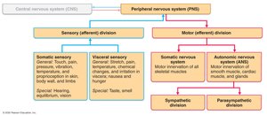
Organization of the PNS
Sensory Receptors, Nerves, Ganglia, and Motor Endings
The PNS consists of sensory receptors, nerves, ganglia, and motor endings. Sensory receptors detect stimuli, nerves are bundles of axons, ganglia are clusters of neuronal cell bodies, and motor endings innervate effectors.
Sensory Receptors: Detect stimuli from internal and external environments.
Nerves: Bundles of peripheral axons transmitting signals.
Ganglia: Clusters of peripheral neuronal cell bodies.
Motor Endings: Axon terminals of motor neurons that innervate muscle fibers and glands.
Peripheral Sensory Receptors
Types and Classification
Sensory receptors are classified by function, location, stimulus type, and structure. They are essential for detecting and transmitting sensory information.
Free Nerve Endings: Monitor general sensory information (pain, temperature).
Complete Receptor Cells: Specialized cells for special sensory information (taste, vision, hearing).
Classification by Location
Exteroceptors: Sensitive to external stimuli; located near body surfaces (touch, pain, temperature).
Interoceptors: Detect internal stimuli; found in viscera (chemical changes, stretching, temperature).
Proprioceptors: Located in muscles, tendons, joints, and ligaments; monitor body movement and stretch.
Classification by Stimulus Type
Mechanoreceptors: Respond to mechanical forces (touch, pressure, vibration, stretch).
Thermoreceptors: Respond to temperature changes.
Chemoreceptors: Respond to chemicals in solution.
Photoreceptors: Respond to light (located in the eye).
Nociceptors: Respond to harmful stimuli resulting in pain.
Classification by Structure
General Sensory Receptors: Widely distributed; monitor touch, pressure, vibration, stretch, pain, temperature, proprioception.
Free Nerve Endings: Abundant in epithelia and connective tissue; respond to pain, temperature, and itch.
Encapsulated Nerve Endings: Mechanoreceptors enclosed in connective tissue; include tactile (Meissner’s), lamellar (Pacinian), bulbous (Ruffini), and proprioceptors.
Proprioceptors
Types and Functions
Proprioceptors are specialized sensory receptors that monitor the position and movement of the body. They are crucial for coordination and balance.
Muscle Spindles: Measure changing length of muscles; contain intrafusal fibers innervated by sensory endings.
Tendon Organs (Golgi): Monitor tension within tendons; important for rapid alternation between flexion and extension.
Joint Kinesthetic Receptors: Located within joint capsules; monitor stretch and send information to the cerebellum and cerebrum.
Cranial Nerves
Overview and Classification
Cranial nerves are twelve pairs of nerves that attach to the brain and pass through specific foramina of the skull. They primarily serve the head and neck, except for the vagus nerve, which extends into the abdomen.
Numbering: Cranial nerves are numbered I–XII.
Attachment: Some attach to the forebrain, others to the brainstem.
Function: Sensory, motor, or mixed functions.
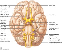
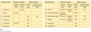
Summary Table of Cranial Nerves
The following table summarizes the sensory and motor functions of the cranial nerves:
Cranial Nerve | Sensory Function | Motor Function |
|---|---|---|
I Olfactory | Special visceral sensory (smell) | - |
II Optic | Special somatic sensory (vision) | - |
III Oculomotor | - | Somatic motor (eye muscles), Visceral motor (pupil, lens) |
IV Trochlear | - | Somatic motor (superior oblique muscle) |
V Trigeminal | General sensory | Somatic motor (chewing muscles) |
VI Abducens | - | Somatic motor (lateral rectus muscle) |
VII Facial | General taste | Somatic motor (facial muscles), Visceral motor (glands) |
VIII Vestibulocochlear | Hearing, equilibrium | - |
IX Glossopharyngeal | General taste | Somatic motor (pharynx), Visceral motor (parotid gland) |
X Vagus | General taste | Somatic motor (pharynx, larynx), Visceral motor (heart, lungs, viscera) |
XI Accessory | - | Somatic motor (trapezius, sternocleidomastoid) |
XII Hypoglossal | - | Somatic motor (tongue muscles) |
Spinal Nerves
Structure and Distribution
Spinal nerves are 31 pairs that connect to the spinal cord and are named according to their point of issue. They are essential for transmitting sensory and motor information between the CNS and the body.
Cervical: 8 pairs (C1–C8)
Thoracic: 12 pairs (T1–T12)
Lumbar: 5 pairs (L1–L5)
Sacral: 5 pairs (S1–S5)
Coccygeal: 1 pair (Co1)
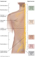
Connection to the Spinal Cord
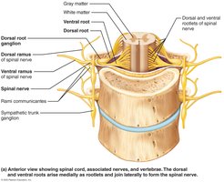
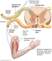
Dorsal Root: Contains sensory fibers; cell bodies in dorsal root ganglion.
Ventral Root: Contains motor fibers from anterior gray column.
Branching and Rami
Dorsal Ramus: Innervates back muscles and skin.
Ventral Ramus: Innervates anterior and lateral trunk, limbs.
Rami Communicantes: Connect to sympathetic chain ganglia.
Nerve Plexuses
Cervical Plexus
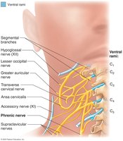
The cervical plexus is formed by the ventral rami of C1–C4 and is located deep to the sternocleidomastoid muscle. It primarily provides cutaneous innervation and some motor innervation to the neck.
Phrenic Nerve: Formed from C3–C5; innervates the diaphragm.
Brachial Plexus
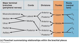
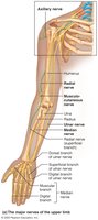
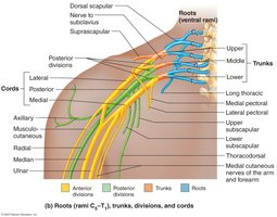
The brachial plexus is formed by ventral rami of C5–T1 and supplies the upper limb. It consists of roots, trunks, divisions, cords, and terminal branches.
Major Nerves: Musculocutaneous, median, ulnar, axillary, radial.
Components: Roots (ventral rami), trunks, divisions, cords.
Lumbar Plexus
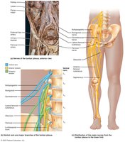
The lumbar plexus arises from L1–L4 and innervates the anterior thigh, abdominal wall, and psoas muscle.
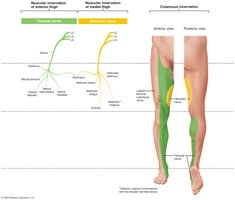
Femoral Nerve: Innervates anterior thigh muscles.
Obturator Nerve: Innervates adductor muscles.
Sacral Plexus
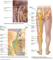
The sacral plexus arises from L4–S4 and is often considered with the lumbar plexus as the lumbosacral plexus. It innervates the lower limb, gluteal region, and perineum.
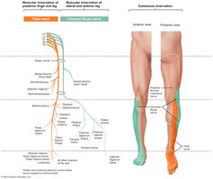
Sciatic Nerve: Largest nerve; consists of tibial and common fibular branches.
Gluteal Nerves: Superior and inferior gluteal nerves innervate gluteal muscles.
Pudendal Nerve: Innervates muscles of the perineum.
Dermatomes
Innervation of the Skin
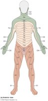
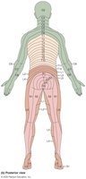
A dermatome is an area of skin innervated by the cutaneous branches of a single spinal nerve. Understanding dermatomes is important for diagnosing nerve injuries and diseases.
Upper Limb: Supplied by spinal nerves of the brachial plexus.
Lower Limb: Anterior surface by lumbar nerves; posterior surface by sacral nerves.
Disorders of the PNS
Common Disorders
Disorders of the PNS can affect sensory and motor function, leading to various clinical symptoms.
Shingles (Herpes Zoster): Viral infection stemming from childhood chickenpox; often triggered by stress and more common in older adults.
Migraine Headache: Related to sensory innervation of cerebral arteries; dilation and irritation of sensory nerve endings cause pain.
Peripheral Neuropathy: Pathological condition of peripheral nerves; symptoms include paresthesia, pain, burning, loss of sensation, muscle weakness, and paralysis. Causes include trauma, repetitive use, systemic disorders (HIV, diabetes, vitamin B deficiency).
