 Indietro
Pulmonary Ventilation and Gas Exchange: The Respiratory System
Guida di studio - Note intelligenti
 Appunti personalizzati basati sui tuoi materiali, ampliati con definizioni chiave, esempi e contesto.
The Respiratory System: Pulmonary Ventilation and Gas Exchange
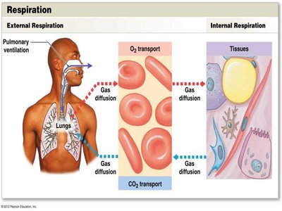
Respiration: External and Internal Processes
Respiration encompasses two integrated processes: external respiration and internal respiration. External respiration involves the exchange of oxygen (O2) and carbon dioxide (CO2) between the body’s interstitial fluids and the external environment, while internal respiration refers to the cellular uptake of O2 and production of CO2 within individual cells.
External Respiration: Gas exchange between alveoli and blood.
Internal Respiration: Gas exchange between blood and tissues.
Pulmonary Ventilation: Physical movement of air in and out of the respiratory tract.
Pulmonary Ventilation: The Mechanics of Breathing
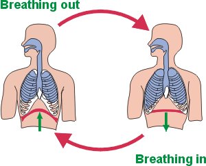
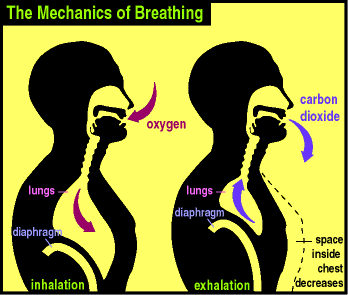
Pulmonary ventilation is the process of moving air into and out of the lungs, primarily to provide alveolar ventilation. This process is driven by changes in thoracic cavity volume, which alter air pressure within the lungs.
Inhalation: Diaphragm contracts and moves downward, increasing thoracic volume and decreasing pressure, drawing air in.
Exhalation: Diaphragm relaxes and moves upward, decreasing thoracic volume and increasing pressure, pushing air out.
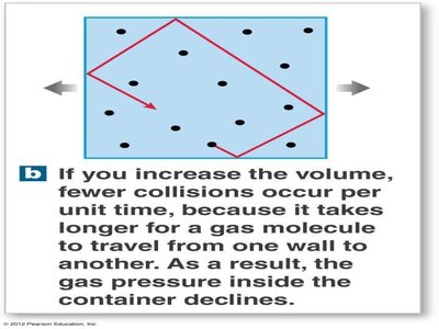
Gas Pressure and Volume: Boyle’s Law
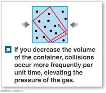
Boyle’s Law defines the relationship between gas pressure (P) and volume (V):
Formula:
If you decrease the volume of a gas, its pressure rises.
If you increase the volume, its pressure decreases.
Pressure and Airflow in the Lungs
Air flows from areas of higher pressure to areas of lower pressure. This principle, along with Boyle’s Law, forms the basis for pulmonary ventilation. The respiratory cycle consists of inspiration (inhalation) and expiration (exhalation).
Volume Changes and Pressure Dynamics
Changes in the volume of the thoracic cavity, due to expansion or contraction of the diaphragm or rib cage, alter the volume of air within the lungs and thus the pressure, facilitating airflow.
Respiratory Cycle: Inhalation and Exhalation
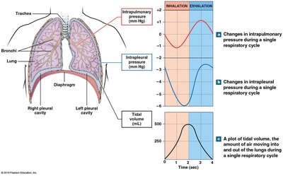
The respiratory cycle is a single cycle of inhalation and exhalation. During inhalation, intrapleural and intrapulmonary pressures fall as the thoracic cavity expands. During exhalation, these pressures rise rapidly, forcing air out as the thoracic cavity volume decreases.
Pneumothorax: Injury to the Chest Wall
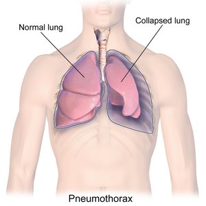
Pneumothorax occurs when air leaks into the space between the lungs and chest wall, breaking the fluid bond between the pleurae and allowing the lung to collapse. Treatment involves removing air from the pleural cavity and sealing the opening to restore normal pressure and allow re-inflation.
Symptoms: Sudden chest pain and shortness of breath.
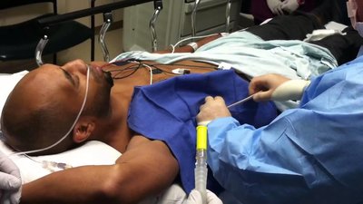
Treatment: Chest tube placement to remove air and restore pressure.
Compliance: Expandability of the Lungs
Compliance refers to the ease with which the lungs can expand. Low compliance requires greater force to fill the lungs, making breathing harder, while high compliance requires less force.
Factors Affecting Compliance
Connective tissue structure: Damage (e.g., emphysema) increases compliance.
Surfactant production: Loss decreases compliance (e.g., respiratory distress syndrome).
Mobility of thoracic cage: Arthritis or skeletal disorders decrease compliance.

The Respiratory Muscles
The diaphragm and external intercostal muscles are the primary muscles involved in normal breathing. Accessory muscles are activated during increased respiratory demand.
Primary muscles: Diaphragm, external intercostals.
Accessory muscles: Sternocleidomastoid, scalenes, pectoralis minor, serratus anterior, internal intercostals, transversus thoracis, abdominal muscles.
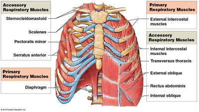
Mechanics of Breathing: Inhalation and Exhalation
Inhalation is always an active process requiring muscle contraction, while exhalation can be passive (muscle relaxation and elastic recoil) or active (muscle contraction during forceful breathing).
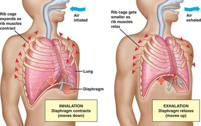
Muscles Used in Normal and Forceful Breathing
Normal Inhalation: Diaphragm (75% of air movement), external intercostals (25%).
Normal Exhalation: Passive process, elastic recoil.
Forceful Inhalation: Accessory muscles (sternocleidomastoid, pectoralis minor, serratus anterior, scalenes).
Forceful Exhalation: Internal intercostals, transversus thoracis, abdominal muscles.
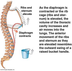
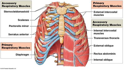
Respiratory Rates and Volumes
The respiratory system adapts to changing oxygen demands by varying respiratory rate and volume. Total lung volume is divided into several volumes and capacities useful for diagnosis.
Resting tidal volume (VT): Amount of air moved in and out during a normal cycle (~500 ml).
Expiratory reserve volume (ERV): Additional air expelled after normal exhalation (males: 1000 ml, females: 700 ml).
Residual volume: Air remaining after maximal exhalation (males: 1200 ml, females: 1100 ml).
Inspiratory reserve volume (IRV): Additional air taken in after normal inhalation (males: 3300 ml, females: 1900 ml).
Calculated Respiratory Capacities
Inspiratory capacity: Tidal volume + inspiratory reserve volume.
Functional residual capacity (FRC): Expiratory reserve volume + residual volume.
Vital capacity: Expiratory reserve volume + tidal volume + inspiratory reserve volume (males: 4800 ml, females: 3400 ml).
Total lung capacity: Vital capacity + residual volume (males: 6000 ml, females: 4200 ml).
Alveolar Ventilation and Dead Space
Only a portion of inhaled air reaches the alveoli for gas exchange. The remainder stays in the conducting passageways and is termed anatomic dead space.
Example: Of 500 ml inhaled, only 350 ml reaches alveoli; 150 ml remains in dead space.
Gas Exchange: Partial Pressures and Diffusion
Gas exchange occurs across the blood-air barrier and depends on partial pressures and diffusion. Dalton’s Law and Henry’s Law govern the movement and solubility of gases.
Dalton’s Law: Each gas in a mixture contributes to total pressure in proportion to its concentration.
Henry’s Law: At a given temperature, the amount of gas dissolved in a liquid is proportional to its partial pressure.
Solubility of Gases in Body Fluids
CO2: Highly soluble in plasma.
O2: Less soluble.
N2: Very low solubility.
Efficiency of Gas Exchange
Large differences in partial pressure across the blood-air barrier.
Short diffusion distances.
Lipid solubility of O2 and CO2.
Large surface area.
Coordinated blood flow and airflow.
Partial Pressures in Alveolar Air and Tissues
Alveolar capillaries: O2 enters blood, CO2 leaves blood.
Tissues: O2 diffuses out of blood, CO2 diffuses into blood.
Oxygen Transport: Hemoglobin and Saturation
O2 binds reversibly to hemoglobin (Hb) in red blood cells, forming oxyhemoglobin (HbO2). Each Hb molecule can bind four O2 molecules.
Hemoglobin saturation: Percentage of heme units with bound O2.
Factors affecting O2 binding: PO2, pH, temperature, metabolic activity.
Oxygen-Hemoglobin Saturation Curve
Higher PO2 results in greater Hb saturation.
Curve shifts right with lower pH or higher temperature (more O2 released).
Curve shifts left with higher pH or lower temperature (less O2 released).
Bohr Effect: Hemoglobin, pH, and CO2
The Bohr effect describes how blood pH affects O2 saturation in Hb. Increased CO2 leads to more carbonic acid, lowering pH and increasing O2 release.
Reaction:
Carbon Dioxide Transport
70% as carbonic acid (H2CO3), dissociates to H+ and HCO3-.
23% bound to hemoglobin (carbaminohemoglobin).
7% dissolved in plasma.
Local and Neural Regulation of Respiration
Peripheral tissues with high PCO2 cause increased blood flow and airflow.
Neural control involves respiratory centers in the medulla oblongata, pons, and cerebral cortex.
Respiratory reflexes are induced by chemoreceptors, baroreceptors, stretch receptors, and other stimuli.
Clinical Considerations: Emphysema and Lung Cancer
Emphysema: Destruction of alveoli reduces surface area for gas exchange, increases compliance, and restricts O2 absorption.
Lung cancer: Aggressive malignancies affecting bronchial passageways or alveoli, often caused by smoking.
Summary Table: Pulmonary Volumes and Capacities
Volume/Capacity | Definition | Male (ml) | Female (ml) |
|---|---|---|---|
Resting Tidal Volume (VT) | Air moved in/out during normal cycle | 500 | 500 |
Expiratory Reserve Volume (ERV) | Additional air expelled after normal exhalation | 1000 | 700 |
Residual Volume | Air remaining after maximal exhalation | 1200 | 1100 |
Inspiratory Reserve Volume (IRV) | Additional air taken in after normal inhalation | 3300 | 1900 |
Vital Capacity | ERV + VT + IRV | 4800 | 3400 |
Total Lung Capacity | Vital capacity + residual volume | 6000 | 4200 |
Example: Spirometry is used to measure these volumes and capacities for diagnostic purposes.
Additional info: These notes expand on brief points with academic context, definitions, and examples to ensure completeness and clarity for exam preparation.
