 Indietro
Reflexes and Reflex Arcs: Structure, Classification, and Examples
Guida di studio - Note intelligenti
 Appunti personalizzati basati sui tuoi materiali, ampliati con definizioni chiave, esempi e contesto.
Reflexes and Reflex Arcs
Overview of Reflexes
Reflexes are rapid, automatic responses to specific stimuli that help maintain homeostasis and protect the body from harm. They are mediated by neural pathways called reflex arcs, which bypass conscious brain processing for speed and efficiency.
Reflex Arc: The neural circuit responsible for a reflex action, typically involving a sensory neuron, interneuron(s), and motor neuron.
Purpose: To provide quick responses to stimuli, often for protection or regulation of physiological functions.
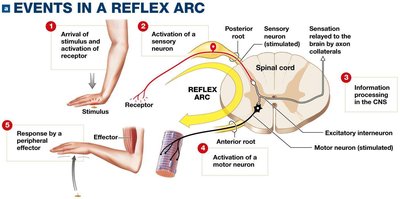
Events in a Reflex Arc
The reflex arc consists of several sequential steps, each involving specific neural structures and functions.
Step 1: Arrival of Stimulus and Activation of Receptor – A stimulus (e.g., touch, pain) activates a sensory receptor.
Step 2: Activation of a Sensory Neuron – The receptor generates an action potential in a sensory neuron.
Step 3: Information Processing in the CNS – The sensory neuron synapses with interneurons in the spinal cord or brain, which process the information.
Step 4: Activation of a Motor Neuron – Interneurons stimulate motor neurons, which carry the response signal.
Step 5: Response by a Peripheral Effector – The motor neuron activates an effector (muscle or gland), producing a response.
Classification of Reflexes
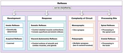
Reflexes can be classified based on development, response, circuit complexity, and processing site.
Development:
Innate Reflexes – Genetically determined, present at birth.
Acquired Reflexes – Learned through experience.
Response:
Somatic Reflexes – Control skeletal muscle contractions.
Visceral (Autonomic) Reflexes – Control actions of smooth and cardiac muscle, and glands.
Complexity of Circuit:
Monosynaptic Reflexes – Involve a single synapse between sensory and motor neuron.
Polysynaptic Reflexes – Involve multiple synapses and interneurons.
Processing Site:
Spinal Reflexes – Processed in the spinal cord.
Cranial Reflexes – Processed in the brain.
Classification | Types |
|---|---|
Development | Innate, Acquired |
Response | Somatic, Visceral (Autonomic) |
Complexity of Circuit | Monosynaptic, Polysynaptic |
Processing Site | Spinal, Cranial |
Monosynaptic Reflexes
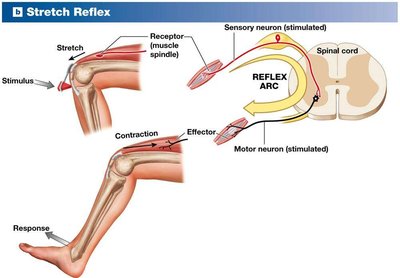
Stretch Reflex
The stretch reflex is a classic example of a monosynaptic reflex, involving only one synapse between the sensory and motor neuron. It helps maintain muscle tone and posture.
Stimulus: Stretching of a muscle (e.g., tapping the patellar tendon).
Receptor: Muscle spindle detects stretch.
Response: Contraction of the stretched muscle.
Muscle Spindle Structure
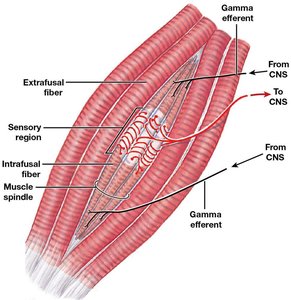
Muscle spindles are specialized sensory receptors within muscles that detect changes in muscle length.
Intrafusal fibers: Specialized muscle fibers within the spindle.
Extrafusal fibers: Regular muscle fibers responsible for contraction.
Sensory region: Area sensitive to stretch.
Gamma efferent: Motor fibers from CNS that adjust spindle sensitivity.
Polysynaptic Reflexes
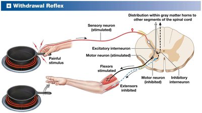
Withdrawal Reflex
The withdrawal reflex is a protective, polysynaptic reflex that removes a body part from a painful stimulus.
Stimulus: Painful stimulus (e.g., touching a hot object).
Pathway: Sensory neuron activates interneurons, which stimulate flexor muscles and inhibit extensors.
Response: Withdrawal of the affected limb.
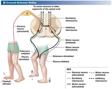
Crossed Extensor Reflex
The crossed extensor reflex complements the withdrawal reflex by activating extensor muscles in the opposite limb to maintain balance.
Stimulus: Painful stimulus to one limb.
Pathway: Sensory neuron activates interneurons that stimulate extensors in the opposite limb and inhibit flexors.
Response: Extension of the opposite limb for support.
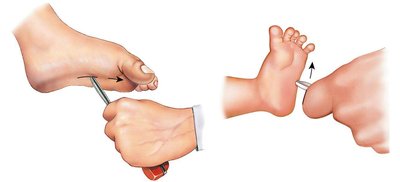
Clinical Example: Babinski Reflex
Babinski Sign
The Babinski reflex is a clinical test for neurological function.
Normal adult response: Toes curl downward.
Infant/abnormal response: Toes fan upward, indicating possible CNS damage.
Summary Table: Reflex Types and Examples
Reflex Type | Example | Key Features |
|---|---|---|
Monosynaptic | Stretch Reflex | One synapse, fast response |
Polysynaptic | Withdrawal Reflex, Crossed Extensor Reflex | Multiple synapses, complex response |
Clinical | Babinski Reflex | Used for neurological assessment |
Key Terms and Concepts
Reflex arc – The neural pathway mediating a reflex.
Muscle spindle – Sensory receptor for stretch reflexes.
Interneuron – Neuron connecting sensory and motor neurons in polysynaptic reflexes.
Effector – Muscle or gland responding to motor neuron stimulation.
Equations and Academic Context
Neural Transmission Equation
The speed of neural transmission can be estimated by: where is velocity, is distance, and is time. Additional info: Reflexes are essential for rapid responses and are used clinically to assess nervous system integrity. The Babinski reflex is particularly important in diagnosing CNS lesions.
