 Indietro
Sensory Pathways and the Somatic Nervous System: Study Notes
Guida di studio - Note intelligenti
 Appunti personalizzati basati sui tuoi materiali, ampliati con definizioni chiave, esempi e contesto.
Sensory Pathways and the Somatic Nervous System
Overview of Sensory and Motor Pathways
The nervous system integrates sensory input and motor output through complex pathways. Sensory pathways transmit information from receptors to the central nervous system (CNS), while motor pathways carry commands from the CNS to effectors. The somatic nervous system specifically controls voluntary movements and conscious sensations.
Afferent Division: Carries sensory information from receptors to the CNS.
Efferent Division: Transmits motor commands from the CNS to effectors (muscles and glands).
Somatic Nervous System: Controls voluntary movements of skeletal muscles.
Signal Transduction and Action Potentials
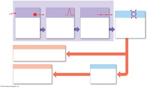
Sensory receptors detect changes in the internal or external environment and convert these stimuli into electrical signals. This process is known as signal transduction.
Sensory Receptor: Structure with nerve endings that responds to a stimulus.
Receptor Potential: Graded potential generated by a sensory receptor in response to a stimulus.
Action Potential: If the receptor potential reaches threshold, an action potential is generated and propagated along the axon to the CNS.
Signal Transduction: Conversion of a receptor potential into an action potential.
Types of Sensory Receptors
General and Special Senses
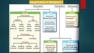
Sensory receptors are classified based on the type of stimulus they detect and their location.
General Senses: Include sensitivity to temperature, pain, touch, pressure, vibration, and proprioception.
Special Senses: Include olfaction (smell), gustation (taste), vision (sight), equilibrium (balance), and hearing. These are provided by specialized receptors located in sense organs.
Classification of Sensory Receptors
Sensory receptors can be classified by their function and location.
Exteroceptors: Detect external stimuli (e.g., touch, temperature).
Proprioceptors: Detect body position and movement (e.g., muscle spindles, Golgi tendon organs).
Interoceptors: Detect internal stimuli (e.g., visceral organs).
Receptive Fields and Sensory Acuity
Receptive Field Concept
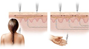
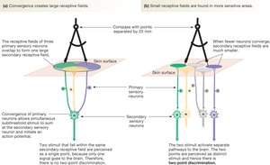
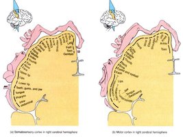
A receptive field is the area in the body that a sensory neuron monitors. The size of the receptive field affects the sensitivity and acuity of perception.
Small Receptive Fields: Found in areas like fingertips and lips; high sensitivity and large representation in the cerebral cortex.
Large Receptive Fields: Found in areas like the back; low sensitivity and small representation in the cortex.
Sensory Receptor Adaptation
Tonic vs. Phasic Receptors
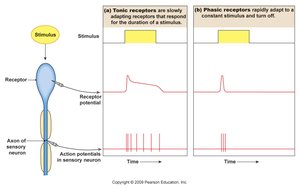
Sensory adaptation refers to the decrease in sensitivity to a continued stimulus. Receptors are classified as tonic or phasic based on their adaptation properties.
Tonic Receptors: Adapt slowly and generate nerve signals steadily (e.g., proprioceptors, nociceptors).
Phasic Receptors: Adapt quickly and generate bursts of action potentials when first stimulated, then reduce or stop signaling (e.g., mechanoreceptors, thermoreceptors).
Types of Tactile Receptors in Skin
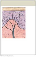
Free Nerve Endings
Free nerve endings are branching tips of sensory neurons that respond to touch, pressure, pain, and temperature.
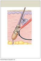
Root Hair Plexus
Root hair plexuses are free nerve endings stimulated by hair movement.
Tactile Discs (Merkel Cells)
Tactile discs are fine touch and pressure receptors sensitive to shape and texture.
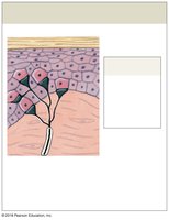
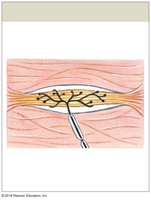
Bulbous Corpuscles (Ruffini)
Bulbous corpuscles are sensitive to pressure and distortion of the deep dermis.
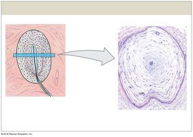
Lamellar Corpuscles (Pacinian)
Lamellar corpuscles are sensitive to deep pressure and high-frequency vibration.
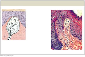
Tactile Corpuscles (Meissner)
Tactile corpuscles are sensitive to fine touch, pressure, and low-frequency vibration.
Baroreceptors and Chemoreceptors
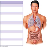
Baroreceptors
Baroreceptors detect changes in pressure within blood vessels and organs, playing a key role in autonomic regulation.
Locations: Carotid sinus, aortic sinus, lungs, digestive tract, colon, bladder wall.
Function: Provide information for reflex control of cardiovascular, respiratory, digestive, and urinary systems.
Chemoreceptors
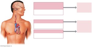
Chemoreceptors are sensitive to changes in pH, CO2, and O2 in blood and cerebrospinal fluid.
Locations: Medulla oblongata, carotid bodies, aortic bodies.
Function: Trigger reflexive adjustments in respiratory and cardiovascular activity.
CNS Coding of Sensory Information
Features of Action Potentials
The CNS distinguishes sensory information based on four features:
Modality: Type of sensation (e.g., smell, taste, touch).
Location: Area where stimulus was applied.
Duration: Length of time stimulus was applied.
Intensity: Strength of stimulus, reflected in the frequency of action potentials.
Key Principle: The magnitude (amplitude) of action potentials is constant; increased stimulus intensity increases the frequency of action potentials.
Somatic Sensory Pathways
Neuronal Organization
Somatic sensory pathways involve three types of neurons:
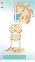
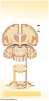
First-order neuron: Delivers sensations to the CNS.
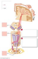
Second-order neuron: Interneuron in the spinal cord or brainstem; crosses to the opposite side (decussation).
Third-order neuron: Located in the thalamus; relays information to the cerebral cortex for awareness.
Ascending Sensory Tracts
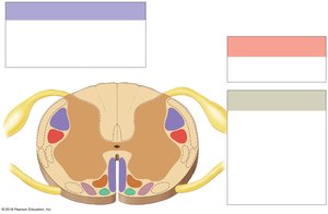
Spinothalamic Pathway: Carries crude touch, pressure, pain, and temperature sensations.
Posterior Column Pathway: Carries highly localized sensations of fine touch, pressure, vibration, and proprioception.
Spinocerebellar Pathway: Carries proprioceptive information to the cerebellum.
Abnormal Sensory Phenomena
Phantom Limb Syndrome: Painful sensations perceived in an amputated limb due to residual activity in severed sensory neurons.
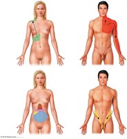
Referred Pain: Pain felt in an area other than the actual source, often due to inappropriate stimulation of secondary sensory neurons.
Somatic Motor Pathways
Descending Motor Tracts
Motor pathways transmit commands from the CNS to skeletal muscles. These pathways are organized into pyramidal and extrapyramidal systems.
Corticospinal (Pyramidal) Pathway: Provides voluntary control over skeletal muscles.
Extrapyramidal Pathways: Control involuntary actions and maintain posture.
Lateral and Medial Pathways: Include rubrospinal, reticulospinal, tectospinal, and vestibulospinal tracts.
Basal Nuclei and Cerebellum
The basal nuclei and cerebellum are responsible for coordination and feedback control over muscle contractions. The cerebellum fine-tunes complex movements through learned patterns and repetition.
Basal Nuclei: Subconscious coordination of movement.
Cerebellum: Conscious fine-tuning and improvement of movement with practice.
Summary Table: Major Sensory and Motor Pathways
Pathway | Function | Key Features |
|---|---|---|
Spinothalamic | Pain, temperature, crude touch | Crosses in spinal cord; awareness in cortex |
Posterior Column | Fine touch, vibration, proprioception | Crosses in medulla; highly localized |
Spinocerebellar | Proprioceptive input | Direct to cerebellum; partial crossing |
Corticospinal | Voluntary motor control | Crosses in brainstem/spinal cord |
Extrapyramidal | Involuntary motor control | Posture, balance, coordination |
Key Equations
Resting Membrane Potential:
Threshold Potential:
Action Potential Frequency:
where f is frequency, N is number of action potentials, and t is time.
Conclusion
The sensory and motor pathways of the somatic nervous system are essential for perceiving the environment and executing voluntary movements. Understanding the organization, adaptation, and coding of sensory information, as well as the structure of motor pathways, is fundamental for students of anatomy and physiology.
