 Indietro
Sensory Receptors and Pathways: General and Special Senses
Guida di studio - Note intelligenti
 Appunti personalizzati basati sui tuoi materiali, ampliati con definizioni chiave, esempi e contesto.
Sensory Receptors and Pathways
Overview of Sensory and Motor Pathways
The nervous system is divided into afferent (sensory) and efferent (motor) divisions, each responsible for transmitting information to and from the central nervous system (CNS). Sensory pathways relay information from receptors to the CNS, while motor pathways control skeletal muscle contractions via the somatic nervous system.
Afferent Division: Includes somatic and visceral sensory pathways.
Efferent Division: Somatic motor portion controls voluntary muscle movements.
Somatic Nervous System (SNS): Controls contractions of skeletal muscles.
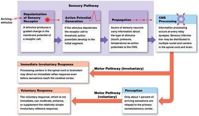
Sensory Receptors: General and Special Senses
Sensory receptors are specialized cells or cell processes that monitor specific conditions in the body or external environment. They are classified into general senses (e.g., touch, pain, temperature) and special senses (e.g., vision, hearing, taste, smell, equilibrium).
General Senses: Include sensitivity to temperature, pain, touch, pressure, vibration, and proprioception.
Special Senses: Include olfaction, gustation, vision, equilibrium, and hearing. These are provided by special sensory receptors located in sense organs and protected by surrounding tissues.
Receptor Specificity and Receptive Fields
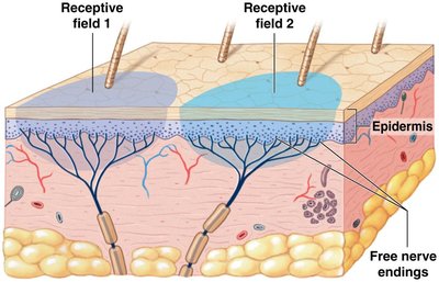
Each sensory receptor has a characteristic sensitivity, known as receptor specificity. The area monitored by a single receptor cell is called its receptive field. The larger the receptive field, the more difficult it is to localize a stimulus.
Receptive Field: Area monitored by a single receptor cell.
Transduction: Conversion of an arriving stimulus into an action potential by a sensory receptor.
Adaptation of Sensory Receptors
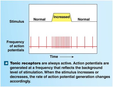
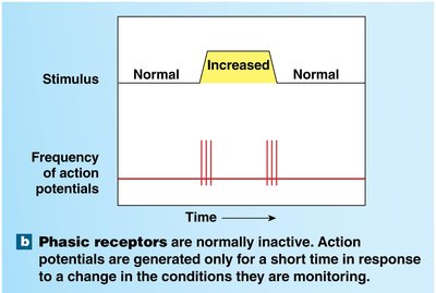
Adaptation refers to the reduction of receptor sensitivity in the presence of a constant stimulus. Peripheral adaptation occurs in the PNS, while central adaptation occurs in the CNS. The nervous system quickly adapts to painless, constant stimuli.
Tonic Receptors: Always active and show little peripheral adaptation. They generate action potentials at a frequency that reflects the background level of stimulation.
Phasic Receptors: Normally inactive and respond strongly at first but then activity decreases. They provide information about the intensity and rate of change of a stimulus.
Types of Sensory Receptors
Nociceptors and Thermoreceptors
Nociceptors are pain receptors found in the superficial skin, joint capsules, periostea of bones, and walls of blood vessels. They do not adapt, serving as reminders of injury or inflammation. Thermoreceptors are temperature receptors located in the dermis, skeletal muscle, liver, and hypothalamus. Cold receptors are more numerous than warm receptors and adapt quickly to stable temperatures.
Chemoreceptors
Chemoreceptors detect small changes in concentrations of specific chemicals such as pH, CO2, and O2. They are important in respiratory centers of the brain and process information unconsciously.
Mechanoreceptors
Mechanoreceptors are sensitive to physical stimuli that distort their plasma membranes. Their membranes contain mechanically gated ion channels that open or close in response to stretching, compression, twisting, or other distortions.
Tactile Receptors: Provide sensations of touch, pressure, and vibration.
Baroreceptors: Detect pressure changes in blood vessels and in digestive, respiratory, and urinary tracts.
Proprioceptors: Monitor positions of joints and skeletal muscles.
Six Types of Tactile Receptors in Skin
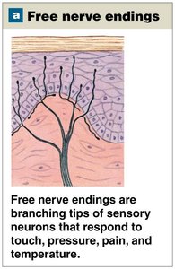
1. Free Nerve Endings
Free nerve endings are sensitive to touch and pressure and are situated between epidermal cells. Tonic receptors with small receptive fields provide touch sensations.
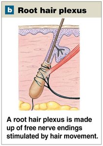
2. Root Hair Plexus Nerve Endings
Root hair plexus nerve endings monitor distortions and movements across the body surface wherever hairs are located. They adapt rapidly and are best at detecting initial contact and subsequent movements.
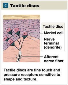
3. Tactile Discs
Tactile discs are fine-touch and pressure receptors sensitive to shape and texture. They are extremely sensitive tonic receptors with very small receptive fields.
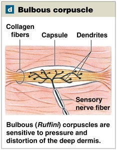
4. Bulbous Corpuscles (Ruffini Corpuscles)
Bulbous corpuscles are sensitive to pressure and distortion of the skin. Located in the reticular (deep) dermis, they are tonic receptors that show little if any adaptation.
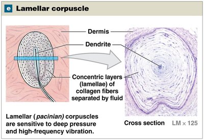
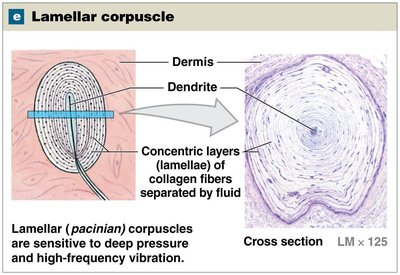
5. Lamellar Corpuscles (Pacinian Corpuscles)
Lamellar corpuscles are sensitive to deep pressure and are fast-adapting receptors. They are most sensitive to pulsing or high-frequency vibrating stimuli. A single dendrite lies within a series of concentric layers of collagen fibers.
6. Tactile Corpuscles (Meissner Corpuscles)
Tactile corpuscles perceive sensations of fine touch, pressure, and low-frequency vibration. They adapt to stimulation within 1 second after contact and are most abundant in eyelids, lips, fingertips, nipples, and external genitalia.
Summary Table: Types of Tactile Receptors
Receptor Type | Location | Stimulus | Adaptation |
|---|---|---|---|
Free Nerve Endings | Epidermis | Touch, pressure, pain, temperature | Tonic |
Root Hair Plexus | Hair follicles | Hair movement | Phasic |
Tactile Discs | Stratum basale | Fine touch, pressure | Tonic |
Bulbous Corpuscles | Deep dermis | Pressure, distortion | Tonic |
Lamellar Corpuscles | Dermis, subcutaneous | Deep pressure, vibration | Phasic |
Tactile Corpuscles | Dermal papillae | Fine touch, low-frequency vibration | Phasic |
Key Concepts and Equations
Action Potential Generation
When a stimulus depolarizes the receptor cell to threshold, action potentials develop in the initial segment. The frequency of action potentials reflects the intensity of the stimulus.
Equation for Action Potential Frequency:
Where f is the frequency, N is the number of action potentials, and t is the time interval.
Adaptation Mechanisms
Peripheral Adaptation: Occurs at the receptor level.
Central Adaptation: Occurs in the CNS.
Applications and Examples
Example: Tactile corpuscles in fingertips allow for precise detection of texture and shape, important for tasks such as reading Braille.
Example: Baroreceptors in blood vessels help regulate blood pressure by detecting changes in vessel stretch.
Additional info: Academic context was added to clarify the function and adaptation of each receptor type, as well as to provide a summary table for comparison.
