 Indietro
Skeletal and Muscular Systems: Structure, Function, and Physiology
Guida di studio - Note intelligenti
 Appunti personalizzati basati sui tuoi materiali, ampliati con definizioni chiave, esempi e contesto.
The Skeletal System
Overview and Functions
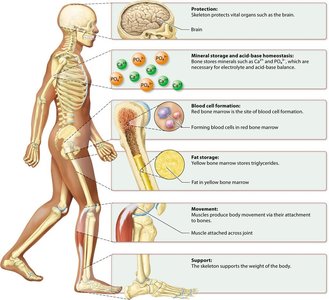
The skeletal system provides the structural framework for the human body, consisting of 206 bones in adults. It serves multiple essential functions:
Protection: Shields vital organs such as the brain, heart, and lungs.
Support: Maintains body shape and supports weight.
Movement: Acts as levers for muscles to produce movement.
Mineral Storage: Stores calcium and phosphate, crucial for homeostasis.
Blood Cell Formation: Red bone marrow produces blood cells (hematopoiesis).
Fat Storage: Yellow bone marrow stores triglycerides.
Bone Structure and Classification
Bones are classified by shape (long, short, flat, irregular, sesamoid) and by tissue type:
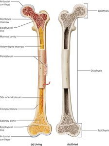
Compact (Cortical) Bone: Dense, organized into osteons (Haversian systems) for strength.
Spongy (Cancellous) Bone: Lattice of trabeculae, found in epiphyses and flat bones, filled with marrow.
Long bones (e.g., femur) have a diaphysis (shaft), epiphyses (ends), medullary cavity (marrow), periosteum (outer membrane), and articular cartilage at joints.
Bone Matrix Composition
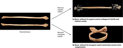
The bone matrix is a composite of organic and inorganic components:
Organic Matrix: Mainly collagen type I, provides flexibility and tensile strength.
Inorganic Matrix: Hydroxyapatite crystals [Ca10(PO4)6(OH)2], confers rigidity and compressive strength.
Removal of either component alters bone properties:
Bone Cells and Remodeling
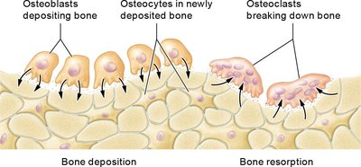
Four main bone cell types regulate bone formation and resorption:
Osteoprogenitor Cells: Stem cells that differentiate into osteoblasts.
Osteoblasts: Synthesize and secrete osteoid, regulate mineralization, and become osteocytes.
Osteocytes: Mature bone cells in lacunae, maintain matrix, act as mechanosensors.
Osteoclasts: Multinucleated cells that resorb bone by secreting acids and enzymes.
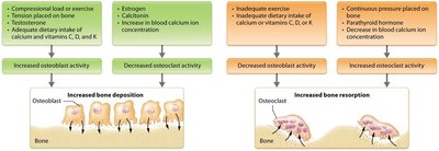
Regulation of Bone Remodeling
Bone remodeling is influenced by mechanical, nutritional, and hormonal factors:
Increased Osteoblast Activity: Compression, exercise, tension, adequate calcium/vitamin intake, estrogen, increased blood calcium.
Increased Osteoclast Activity: Continuous pressure, parathyroid hormone, decreased blood calcium.
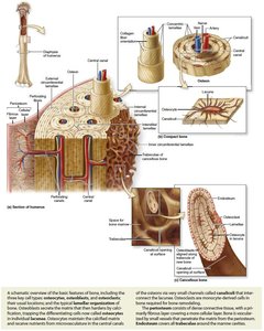
Histology of Compact Bone
Compact bone is organized into osteons, each consisting of concentric lamellae around a central canal (Haversian canal) containing blood vessels and nerves. Osteocytes reside in lacunae and communicate via canaliculi.
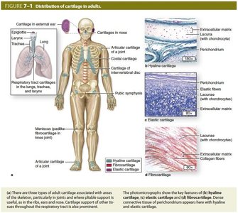
Cartilage Types and Distribution
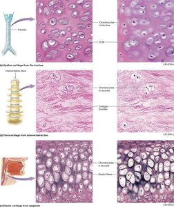
Cartilage is a resilient connective tissue formed by chondroblasts and maintained by chondrocytes in lacunae. Three types exist:
Hyaline Cartilage: Most common, found in joints, nose, trachea; provides support and flexibility.
Elastic Cartilage: Contains elastic fibers, found in ear and epiglottis; highly flexible.
Fibrocartilage: Dense collagen, found in intervertebral discs and pubic symphysis; resists compression.
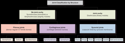
Classification of Joints
Joints are classified by structure and function:
Fibrous Joints: Bones united by fibrous tissue; mostly immovable (e.g., sutures).
Cartilaginous Joints: Bones united by cartilage; slightly movable (e.g., symphysis).
Synovial Joints: Freely movable, with a fluid-filled cavity (e.g., knee, shoulder).
The Muscular System
Overview and Muscle Types
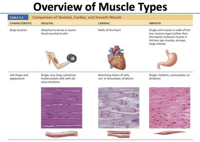
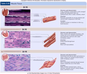
Muscle tissue enables movement, posture, and vital functions. There are three types:
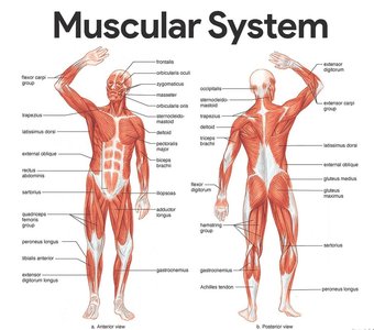
Skeletal Muscle: Voluntary, striated, attached to bones.
Cardiac Muscle: Involuntary, striated, found in the heart.
Smooth Muscle: Involuntary, non-striated, found in walls of hollow organs.
Skeletal Muscle Structure
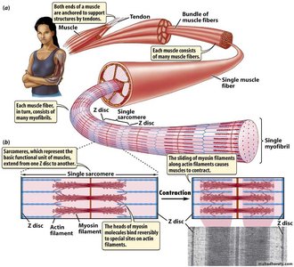
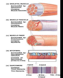
Skeletal muscle is organized hierarchically:
Whole Muscle: Enclosed by epimysium.
Fascicles: Bundles of fibers, surrounded by perimysium.
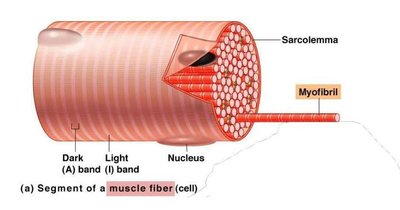
Muscle Fiber (Cell): Multinucleate, surrounded by endomysium.
Myofibrils: Contractile units within fibers.
Sarcomere: Functional unit, bounded by Z-lines, contains actin and myosin filaments.
Sliding Filament Theory
Muscle contraction occurs as actin filaments slide past myosin filaments, shortening the sarcomere. The cross-bridge cycle involves:
Calcium binding to troponin, shifting tropomyosin and exposing actin sites.
Myosin heads bind actin, perform a power stroke, release ADP, detach with new ATP, and re-cock.
Neuromuscular Junction and Excitation-Contraction Coupling
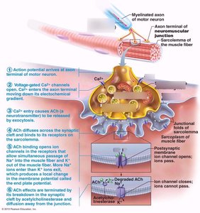
The neuromuscular junction (NMJ) is where a motor neuron communicates with a muscle fiber:
Action potential triggers Ca2+ influx, acetylcholine release, and binding to nicotinic receptors.
End plate potential propagates, leading to Ca2+ release from the sarcoplasmic reticulum and contraction.
Acetylcholinesterase terminates the signal by degrading acetylcholine.
Metabolism, Cellular Respiration, and ATP
Metabolism Overview
Metabolism includes catabolism (breakdown for ATP production) and anabolism (synthesis using ATP). Key pathways:
Glycolysis: Glucose breakdown in cytosol, yields 2 ATP and 2 NADH per glucose.
Citric Acid Cycle: In mitochondria, produces NADH, FADH2, GTP/ATP, and CO2.
Electron Transport Chain: Generates a proton gradient for ATP synthesis; oxygen is the final electron acceptor.
Beta Oxidation: Fatty acids broken into acetyl-CoA for ATP production.
Amino Acid Metabolism: Amino acids enter energy pathways during starvation.
ATP is the primary energy currency, powering transport, contraction, and biosynthesis.
Caloric Values: Carbohydrates and protein: 4 kcal/g; fat: 9 kcal/g; alcohol: 7 kcal/g; water: 0 kcal/g.
Tables
Major Bones — Location and Clinical Correlation
Bone | Skeleton Division | Anatomical Location | Clinical Correlation |
|---|---|---|---|
Temporal Bone | Axial (Cranium) | Lateral skull; houses middle & inner ear | Fracture can damage facial nerve (CN VII) and cochlea |
Sternum | Axial (Thorax) | Anterior midline of chest | Site of bone marrow biopsy; CPR compressions |
Clavicle | Appendicular (Pectoral Girdle) | Between manubrium and scapula | Most commonly fractured bone; falls on outstretched hand |
Humerus | Appendicular (Upper Limb) | Arm | Radial nerve injury with spiral groove fracture causes wrist drop |
Radius | Appendicular (Upper Limb) | Lateral forearm | Colles' fracture (distal radius) — common in falls |
Femur | Appendicular (Lower Limb) | Thigh | Hip fracture (femoral neck) — major cause of morbidity in elderly |
Tibia | Appendicular (Lower Limb) | Medial leg | Stress fractures common in runners; shin splints involve periosteum |
Vertebrae (L4–L5) | Axial (Vertebral Column) | Lumbar spine | Common site of herniated disc; lumbar puncture at L4–L5 |
Major Skeletal Muscles — Origin, Insertion, Action, and Innervation
Muscle | Origin | Insertion | Primary Action | Innervation |
|---|---|---|---|---|
Deltoid | Clavicle; acromion; scapular spine | Deltoid tuberosity of humerus | Arm abduction, flexion, extension | Axillary nerve (C5, C6) |
Biceps Brachii | Coracoid process; supraglenoid tubercle | Radial tuberosity; bicipital aponeurosis | Forearm flexion; supination | Musculocutaneous nerve (C5, C6) |
Triceps Brachii | Infraglenoid tubercle; posterior humerus | Olecranon process of ulna | Forearm extension | Radial nerve (C6, C7, C8) |
Pectoralis Major | Clavicle; sternum; costal cartilages 1–6 | Intertubercular groove of humerus | Arm flexion, adduction, medial rotation | Medial and lateral pectoral nerves (C5–T1) |
Latissimus Dorsi | Spinous processes T7–T12; thoracolumbar fascia; iliac crest; lower ribs | Intertubercular groove of humerus | Arm extension, adduction, medial rotation | Thoracodorsal nerve (C6, C7, C8) |
Rectus Abdominis | Pubic symphysis; pubic crest | Xiphoid process; costal cartilages 5–7 | Trunk flexion; compression of abdomen | Intercostal nerves T7–T12 |
Iliopsoas | Iliac fossa (iliacus); lumbar vertebral bodies (psoas) | Lesser trochanter of femur | Hip flexion; thigh medial rotation | Femoral nerve (L2, L3, L4) |
Gluteus Maximus | Posterior iliac crest; sacrum; coccyx | Gluteal tuberosity; iliotibial tract | Hip extension; lateral rotation | Inferior gluteal nerve (L5, S1, S2) |
Quadriceps Femoris | ASIS; anterior iliac spine; anterior femur (three vasti) | Tibial tuberosity via patellar tendon | Knee extension; hip flexion (rectus femoris) | Femoral nerve (L2, L3, L4) |
Gastrocnemius | Medial and lateral femoral condyles | Calcaneal (Achilles) tendon → calcaneus | Plantarflexion of foot; knee flexion | Tibial nerve (S1, S2) |
Key Terminology
Osteoblast: Bone-forming cell that synthesizes osteoid and regulates mineralization.
Osteoclast: Bone-resorbing cell that dissolves mineral and degrades matrix.
Osteocyte: Mature bone cell in lacunae, mechanosensor.
Periosteum: Membrane covering bone, essential for growth and repair.
Synovial Joint: Freely movable joint with a cavity lined by synovial membrane.
Osteoporosis: Disease with reduced bone density and increased fracture risk.
Sarcomere: Contractile unit of muscle, site of cross-bridge cycling.
Sliding Filament Theory: Mechanism of muscle contraction via actin-myosin interaction.
Neuromuscular Junction: Synapse between motor neuron and muscle fiber.
Motor Unit: Motor neuron and all fibers it innervates.
Acetylcholinesterase: Enzyme terminating acetylcholine action at NMJ.
Myasthenia Gravis: Autoimmune disorder affecting NMJ.
Key Equations
Glucose oxidation:
Additional info:
Some explanations and context were expanded for clarity and completeness, including clinical correlations and metabolic pathways.
