 Indietro
Skeletal and Smooth Muscle: Structure, Function, and Disorders
Guida di studio - Note intelligenti
 Appunti personalizzati basati sui tuoi materiali, ampliati con definizioni chiave, esempi e contesto.
Muscle Tissue Types and Their Characteristics
Overview of Muscle Tissue
Muscle tissue is essential for movement, stability, and various physiological functions. The three main types of muscle tissue are skeletal muscle, cardiac muscle, and smooth muscle. Each type has unique structural and functional properties.
Comparison of Skeletal, Cardiac, and Smooth Muscle
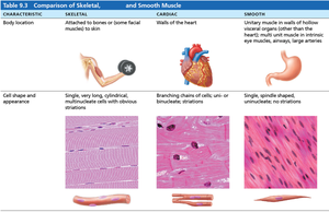
The following table summarizes the key differences among skeletal, cardiac, and smooth muscle tissues:
Characteristic | Skeletal | Cardiac | Smooth |
|---|---|---|---|
Body Location | Attached to bones or skin | Walls of the heart | Walls of hollow organs (except heart) |
Cell Shape & Appearance | Long, cylindrical, multinucleate, striated | Branching, uni- or binucleate, striated | Spindle-shaped, uninucleate, no striations |
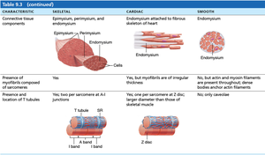
Connective Tissue Components | Epimysium, perimysium, endomysium | Endomysium attached to fibrous skeleton | Endomysium only |
Presence of Myofibrils | Yes | Yes, irregular thickness | No, but actin and myosin filaments present |
Presence & Location of T Tubules | Yes, at A-I junctions | Yes, at Z discs | Only caveolae |
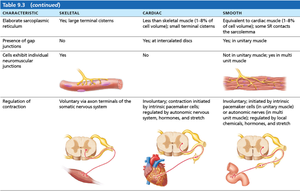
Elaborate Sarcoplasmic Reticulum | Yes, large terminal cisterns | Less than skeletal muscle | Equivalent to cardiac muscle |
Presence of Gap Junctions | No | Yes, at intercalated discs | Yes, in unitary muscle |
Cells Exhibit Individual Neuromuscular Junctions | Yes | No | No (except in multi unit muscle) |
Regulation of Contraction | Voluntary (somatic nervous system) | Involuntary (autonomic nervous system) | Involuntary (autonomic nervous system) |
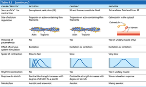
Source of Ca2+ for Contraction | Sarcoplasmic reticulum | SR and extracellular fluid | Extracellular fluid and SR |
Site of Calcium Regulation | Troponin on actin | Troponin on actin | Calmodulin in cytosol |
Speed of Contraction | Slow to fast | Slow | Very slow |
Rhythmic Contraction | No | Yes | Yes (in unitary muscle) |
Metabolism | Aerobic and anaerobic | Aerobic | Mainly aerobic |
Smooth Muscle Structure and Function
Organization of Smooth Muscle
Smooth muscle is found in the walls of most hollow organs, such as the respiratory, digestive, urinary, and reproductive tracts, as well as blood vessels (except the heart). It is typically organized into two layers:
Longitudinal layer: Fibers run parallel to the organ's long axis; contraction shortens the organ.
Circular layer: Fibers run around the circumference; contraction constricts the lumen.
Alternating contractions and relaxations mix and propel substances through the organ's lumen.
Myofilament Arrangement in Smooth vs. Skeletal Muscle
Smooth muscle fibers differ from skeletal muscle fibers in several ways:
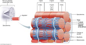
Smooth muscle has less elaborate sarcoplasmic reticulum (SR) and lacks T tubules.
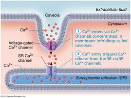
SR stores some Ca2+, but most calcium for contraction comes from extracellular sources.
The sarcolemma contains caveolae, pouchlike infoldings with Ca2+ channels for rapid influx.
Skeletal muscle has a well-developed SR and T tubules, storing all intracellular Ca2+ for contraction.
Disorders of the Muscular System
Duchenne Muscular Dystrophy (DMD)
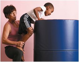
Duchenne muscular dystrophy is a severe, inherited muscle-destroying disease, primarily affecting males. It is caused by a defective gene for dystrophin, a protein that stabilizes the sarcolemma. The disease typically appears in early childhood and progresses from the extremities to the head, chest, and cardiac muscles.
Symptoms: Clumsiness, frequent falls, progressive muscle weakness.
Pathophysiology: Sarcolemma tears easily, allowing excess Ca2+ entry, damaging contractile fibers.
Inflammation and loss of regenerative capacity lead to increased apoptosis and muscle mass loss.
Supportive care can extend life into the 30s and beyond.
Myasthenia Gravis
Myasthenia gravis is an autoimmune disease characterized by muscle weakness, drooping eyelids, and difficulty swallowing and talking. It results from a shortage of acetylcholine (ACh) receptors, which are attacked by the body's own antibodies.
Symptoms: Drooping eyelids, difficulty swallowing/talking, generalized muscle weakness.
Pathophysiology: Fewer ACh receptors at neuromuscular junctions, impaired muscle activation.
Rigor Mortis
Rigor mortis is the stiffening of muscles after death, beginning 3–4 hours postmortem and peaking at 12 hours. It occurs because ATP production ceases, preventing Ca2+ reuptake and cross bridge detachment, resulting in sustained contraction until proteins break down.
Intracellular Ca2+ increases, cross bridges form and cannot detach.
Muscles remain contracted until protein degradation releases myosin from actin.
Disuse Atrophy
Disuse atrophy is the degeneration and loss of muscle mass due to inactivity or loss of neural stimulation. It can begin almost immediately, with muscle strength declining by 5% per day. Paralyzed muscles may shrink to one-fourth their original size, and lost muscle tissue is replaced by fibrous connective tissue, making rehabilitation impossible.
Disorders of the Nervous System
Multiple Sclerosis and Neurotoxins
Disorders affecting nervous tissue can lead to symptoms such as visual disturbances, weakness, loss of muscular control, speech disturbances, and incontinence. Multiple sclerosis is a demyelinating disorder treated with drugs that modify immune system activity. Maintaining high vitamin D levels may reduce risk.
Clinical Reference: Neurotoxins and Their Effects
Various toxins and drugs affect nervous tissue and neuromuscular function:
Novocaine: Blocks voltage-gated Na+ channels.
Curare: Blocks chemically-gated Na+ channels.
Tetrodotoxin & Saxitoxin: Block Na+ channels.
Batrachotoxin & DDT: Prevent closure of Na+ channels.
a-Bungarotoxin: Blocks neurotransmitter release.
Scorpion venom: Potentiates ACh release.
Black widow spider venom: Causes explosive neurotransmitter release.
Botulinum toxin: Blocks ACh release at NMJ.
Atropine: Blocks ACh receptors.
Nerve gas, Organophosphates: Inhibit acetylcholinesterase.
Nicotine: Binds to cholinergic receptors.
Strychnine: Inhibits glycine at CNS post-synaptic sites, increasing excitability.
Key Terms and Concepts
Myofilaments: Actin and myosin filaments responsible for muscle contraction.
Sarcoplasmic Reticulum (SR): Organelle storing Ca2+ for contraction.
Caveolae: Infoldings in smooth muscle sarcolemma containing Ca2+ channels.
Neuromuscular Junction: Synapse between motor neuron and muscle fiber.
Dystrophin: Protein stabilizing muscle cell membrane.
Acetylcholine (ACh): Neurotransmitter for muscle activation.
Relevant Equations
Muscle contraction depends on calcium ion concentration and ATP availability:
Calcium Regulation:
ATP Requirement for Cross Bridge Detachment:
Muscle Atrophy Rate:
