 Indietro
Skeletal Muscle Anatomy: Structure and Organization
Guida di studio - Note intelligenti
 Appunti personalizzati basati sui tuoi materiali, ampliati con definizioni chiave, esempi e contesto.
Skeletal Muscle Anatomy
Overview of Skeletal Muscle Structure
Skeletal muscle is a complex organ composed of multiple tissue types, including muscle fibers, nerves, blood vessels, and connective tissues. Understanding its hierarchical organization is essential for grasping muscle function and physiology.
Skeletal muscle is a discrete organ, made up of bundles of muscle fibers.
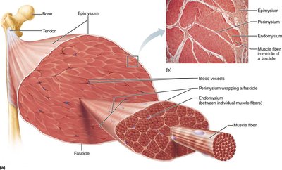
Each muscle is served by nerves, arteries, and veins, which branch extensively within connective tissue sheaths.
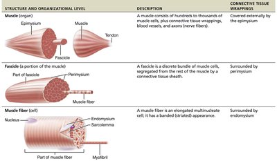
Connective tissue sheaths include the epimysium, perimysium, and endomysium.
Connective Tissue Sheaths
Connective tissue sheaths play a critical role in supporting and organizing muscle fibers:
Epimysium: Surrounds the entire muscle.
Perimysium: Surrounds each fascicle (bundle of muscle fibers).
Endomysium: Surrounds individual muscle fibers.
Muscle Fiber Structure
Characteristics of Muscle Fibers
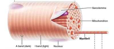
Each skeletal muscle fiber is a long, cylindrical cell with multiple nuclei located just beneath the sarcolemma (plasma membrane). Muscle fibers contain specialized structures for contraction and energy storage.
Diameter: 10–100 µm; Length: up to 30 cm.
Sarcoplasm: Cytoplasm of the muscle cell, containing glycosomes (glycogen storage) and myoglobin (oxygen storage).
Contains organelles such as mitochondria, myofibrils, sarcoplasmic reticulum, and T tubules.
Myofibrils and Sarcomeres
Myofibrils
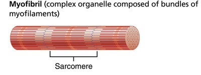
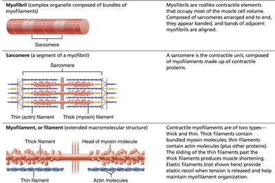
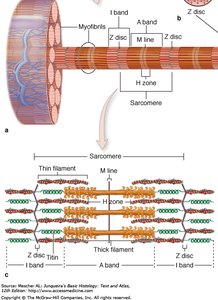
Myofibrils are rodlike contractile elements that occupy most of the muscle cell volume. They are composed of repeating units called sarcomeres.
Each muscle fiber contains hundreds to thousands of myofibrils.
Myofibrils are densely packed, accounting for about 80% of cellular volume.
Sarcomere: The Functional Unit
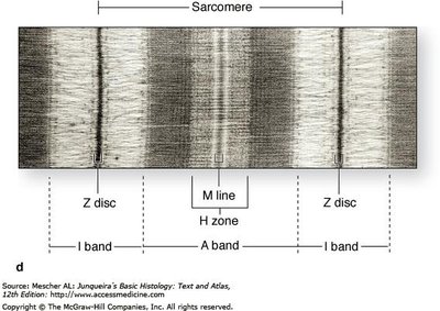
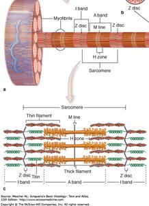
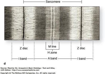
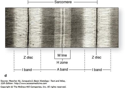
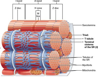
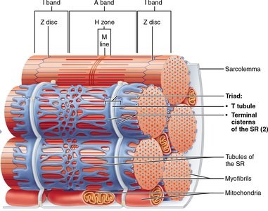
The sarcomere is the smallest contractile unit of a muscle fiber. It is defined as the region between two successive Z discs and contains an A band flanked by half an I band at each end.
A band: Dark band, contains thick filaments (myosin).
I band: Light band, contains thin filaments (actin).
Z disc: Bisects the I band, anchors thin filaments.
H zone: Lighter region in the center of the A band, only thick filaments present.
M line: Bisects the H zone, connects adjacent thick filaments.
Myofilaments: Thick and Thin Filaments
Types of Myofilaments
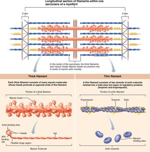
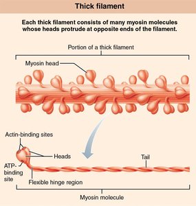
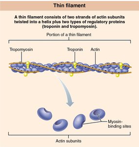
Myofilaments are the contractile proteins within the sarcomere. There are two main types:
Thick filaments: Composed primarily of myosin.
Thin filaments: Composed primarily of actin, along with regulatory proteins troponin and tropomyosin.
Sarcoplasmic Reticulum and T Tubules
Sarcoplasmic Reticulum (SR)
The sarcoplasmic reticulum is an elaborate smooth endoplasmic reticulum that regulates intracellular calcium levels. It stores calcium and releases it on demand during muscle contraction.
Calcium release from the SR is essential for muscle contraction.
T Tubules
T tubules are extensions of the sarcolemma that penetrate deep into the muscle fiber at each A band–I band junction. They help transmit the action potential to the interior of the muscle fiber, ensuring coordinated contraction.
The lumen of the T tubule is continuous with the extracellular space.
Summary Table: Skeletal Muscle Structure
Structure and Organizational Level | Description | Connective Tissue Wrappings |
|---|---|---|
Muscle (organ) | A muscle consists of hundreds to thousands of muscle cells, plus connective tissue wrapping, blood vessels, and nerves (nerve fiber). | Covered externally by the epimysium |
Fascicle (portion of the muscle) | A fascicle is a discrete bundle of muscle cells, segregated from the rest of the muscle by a connective tissue sheath. | Surrounded by perimysium |
Muscle fiber (cell) | A muscle fiber is an elongated multinucleate cell; it has a banded (striated) appearance. | Surrounded by endomysium |
Key Terms and Definitions
Epimysium: Outer connective tissue covering the entire muscle.
Perimysium: Connective tissue surrounding each fascicle.
Endomysium: Connective tissue surrounding each muscle fiber.
Sarcolemma: Plasma membrane of a muscle cell.
Sarcoplasm: Cytoplasm of a muscle cell.
Myofibril: Rodlike contractile element within muscle fibers.
Sarcomere: Smallest contractile unit of muscle, defined by Z discs.
Myofilament: Protein filaments (thick and thin) within the sarcomere.
Sarcoplasmic reticulum: Specialized endoplasmic reticulum for calcium storage.
T tubule: Extension of the sarcolemma into the muscle fiber.
Example: Muscle Contraction
During muscle contraction, the sarcoplasmic reticulum releases calcium ions, which bind to troponin on the thin filaments. This allows myosin heads on the thick filaments to bind to actin, resulting in the sliding of filaments and shortening of the sarcomere.
Relevant Equations
Muscle force is proportional to the number of cross-bridges formed between actin and myosin:
Where: = total muscle force = number of cross-bridges = force per cross-bridge
*Additional info: The notes have been expanded to provide academic context and definitions for clarity and completeness.*
