 Indietro
Skeletal Muscle Contraction: Structure, Physiology, and Mechanisms
Guida di studio - Note intelligenti
 Appunti personalizzati basati sui tuoi materiali, ampliati con definizioni chiave, esempi e contesto.
Muscle Tissue Structure and Organization
Skeletal Muscle Fiber Anatomy
Skeletal muscle fibers are highly organized cells specialized for contraction. Their internal structure is composed of repeating units called sarcomeres, which are the fundamental contractile units. - Sarcolemma: The plasma membrane of the muscle fiber. - Myofibrils: Cylindrical structures within the muscle fiber, composed of sarcomeres. - Sarcoplasmic Reticulum (SR): Specialized endoplasmic reticulum that stores calcium ions. - T Tubules: Invaginations of the sarcolemma that help transmit action potentials deep into the muscle fiber. 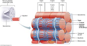
Sarcomere Structure
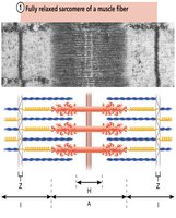
The sarcomere is defined by Z discs and contains thick (myosin) and thin (actin) filaments. - Z disc: Boundary of the sarcomere. - M line: Center of the sarcomere. - Thick filaments: Composed of myosin. - Thin filaments: Composed of actin, troponin, and tropomyosin. 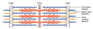
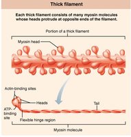
Thin and Thick Filaments
- Thin Filament: Consists of actin subunits, troponin, and tropomyosin. Troponin and tropomyosin regulate access to myosin-binding sites on actin. 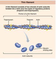 - Thick Filament: Composed of myosin molecules, each with a head that binds actin and ATP.
Sliding Filament Mechanism of Muscle Contraction
Overview of the Sliding Filament Model
Muscle contraction occurs when thin filaments slide past thick filaments, shortening the sarcomere. This process is regulated by calcium ions and the proteins troponin and tropomyosin. - Troponin: Binds calcium, causing a conformational change. - Tropomyosin: Moves away from myosin-binding sites on actin when troponin binds calcium. - Actin: Provides binding sites for myosin heads. 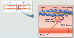
Muscle Fiber Contraction: Cross Bridge Cycling
Steps of Cross Bridge Cycle
The cross bridge cycle is the repeated sequence of events that causes muscle contraction. 1. Cross-bridge formation: Energized myosin head binds to actin. 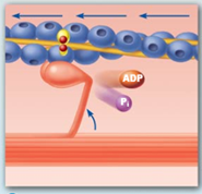 2. Power stroke: Myosin head pivots, pulling actin toward the M line; ADP and Pi are released.
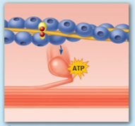 3. Cross-bridge detachment: ATP binds to myosin, causing it to detach from actin.
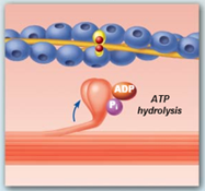 4. Cocking of myosin head: ATP is hydrolyzed to ADP and Pi, re-energizing the myosin head.
 Example: During muscle contraction, these steps repeat as long as calcium and ATP are available.
Changes in Sarcomere During Contraction
Relaxed vs. Contracted Sarcomere
- In a relaxed muscle, the sarcomere is at its maximum length. - During contraction, the sarcomere shortens as thin filaments slide inward. 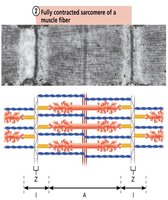
Neuromuscular Junction and Muscle Fiber Excitation
Structure of the Neuromuscular Junction
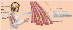
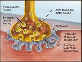
The neuromuscular junction is the synapse between a motor neuron and a skeletal muscle fiber. - Axon terminal: Releases acetylcholine (ACh) into the synaptic cleft. - Synaptic cleft: Space between neuron and muscle fiber. - Junctional folds: Increase surface area for ACh receptors. 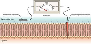
Role of Acetylcholine (ACh)
- ACh is released from the motor neuron and binds to receptors on the sarcolemma, triggering an action potential in the muscle fiber.
Resting Membrane Potential and Ion Channels
Resting Membrane Potential
The resting membrane potential is the voltage difference across the sarcolemma when the muscle cell is at rest, typically around -70 mV. - Reference electrode: Measures extracellular fluid. - Recording microelectrode: Measures cytosol. 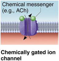
Types of Ion Channels
- Chemically gated ion channels: Open in response to binding of a chemical messenger (e.g., ACh). 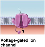 - Voltage-gated ion channels: Open in response to changes in membrane potential.
Excitation-Contraction Coupling
Sequence of Events
Excitation-contraction coupling links the action potential in the sarcolemma to muscle contraction. - Action potential travels along sarcolemma and into T tubules. - Voltage-sensitive proteins trigger Ca2+ release from the sarcoplasmic reticulum. - Ca2+ binds to troponin, allowing cross bridge cycling. Equation:
Muscle Fiber Relaxation
Role of ATP and Myosin ATPase
- ATP is required for detachment of myosin from actin and for re-cocking the myosin head. - Myosin ATPase hydrolyzes ATP to provide energy for contraction. - In the absence of ATP, muscles enter a rigor state (rigor mortis).
Types of Skeletal Muscle Fibers
Comparison of Fiber Types
Skeletal muscle fibers are classified based on their anatomical and metabolic properties.
Type | Characteristics | Metabolism |
|---|---|---|
Slow Oxidative (Type I) | Small diameter, high endurance, rich in mitochondria | Aerobic |
Fast Oxidative (Type IIa) | Intermediate diameter, moderate endurance | Aerobic & Anaerobic |
Fast Glycolytic (Type IIb/IIx) | Large diameter, low endurance, high power | Anaerobic |
Example: Marathon runners have more slow oxidative fibers, while sprinters have more fast glycolytic fibers.
Summary: Steps in Skeletal Muscle Contraction
Four main steps must occur for skeletal muscle contraction: 1. Events at the neuromuscular junction 2. Muscle fiber excitation 3. Excitation-contraction coupling 4. Cross bridge cycling Additional info: These steps are tightly regulated and require precise coordination of electrical and chemical signals. ----------------------------------------
