 Indietro
Skeletal Muscle Groups, Structure, and Sarcomere Organization
Guida di studio - Note intelligenti
 Appunti personalizzati basati sui tuoi materiali, ampliati con definizioni chiave, esempi e contesto.
Muscle Groups and Their Functions
Major Skeletal Muscle Groups
Skeletal muscles are organized into functional groups that perform specific movements. Understanding these groups and their constituent muscles is essential for studying human movement and anatomy.
Quadriceps (Front of thigh):
Rectus femoris
Vastus lateralis
Vastus medialis
Vastus intermedius
Main Function: Knee extension. Study Tip: Remember "Vastus" makes up three of them, and "Rectus" runs down the center.
Hamstrings (Back of thigh):
Biceps femoris
Semitendinosus
Semimembranosus
Main Function: Knee flexion & hip extension. Study Tip: The two "Semi" muscles are medial, Biceps femoris is lateral.
Rotator Cuff (Shoulder stabilizer):
Supraspinatus
Infraspinatus
Teres minor
Subscapularis
Main Function: Shoulder stabilization (Glenohumeral joint). Study Tip: Use the acronym SITS to memorize these four.
Erector Spinae (Lower/mid back):
Iliocostalis
Longissimus
Spinalis
Main Function: Spine extension & posture. Study Tip: Use the acronym I Love Spine (Iliocostalis, Longissimus, Spinalis) ordered from lateral to medial.
Calves (Lower leg):
Gastrocnemius
Soleus
Plantaris*
Main Function: Plantar flexion (pointing toes). Study Tip: Gastrocnemius is the visible, two-headed outer muscle; soleus sits flat underneath. *Triceps Surae does not include the Plantaris.
Extraocular Muscles & Movements
These muscles control eye movement and direction:
Muscle Name | Primary Action | Visual Direction |
|---|---|---|
Lateral Rectus | Rotates eyeball laterally | Outward / Laterally (away from the nose) |
Medial Rectus | Rotates eyeball medially | Inward / Medially (toward the nose) |
Superior Rectus | Rotates eyeball superiorly | Upward (and slightly inward) |
Inferior Rectus | Rotates eyeball inferiorly | Downward (and slightly inward) |
Superior Oblique | Rotates eyeball inferiorly & laterally | Downward and Outward |
Inferior Oblique | Rotates eyeball superiorly & laterally | Upward and Outward |
Other Muscle Groups and Actions
Muscles are classified by their actions at joints:
Hip/Thigh Abduction: Gluteus Maximus, Gluteus Medius, Gluteus Minimus, Tensor fascia latae, Sartorius (weak), Piriformis (only when bent)
Hip/Thigh Adduction: Gracilis, Adductor Longus, Pectineus
Hip/Thigh Lateral Rotation: Gluteus Maximus, Sartorius, Piriformis
Hip/Thigh Medial Rotation: Gluteus Medius, Gluteus Minimus, Pectineus, Adductor Longus, Tensor fascia Latae, Gracilis
Hip/Thigh Extension: Gluteus Maximus, Biceps Femoris, Semimembranosus, Semitendinosus
Hip/Thigh Flexion: Rectus Femoris, Sartorius, Pectineus, Adductor Longus, Tensor Fascia Latae
Knee Extension: Rectus femoris, Vastus Lateralis, Vastus Medialis, Vastus Intermedius
Knee Flexion: Biceps Femoris, Semimembranosus, Semitendinosus, Sartorius*, Gracilis*, Gastrocnemius*, Popliteus*
Dorsiflexion: Tibialis Anterior, Extensor Digitorum, Extensor Hallucis Longus
Plantarflexion: Fibularis Longus, Fibularis Brevis, Gastrocnemius, Soleus, Plantaris, Tibialis Posterior, Flexor Digitorum Longus, Flexor Hallucis Longus
Inversion (medial): Tibialis Anterior, Tibialis Posterior, Flexor Digitorum Longus, Flexor Hallucis Longus
Eversion (lateral): Fibularis Longus, Fibularis Brevis
Master List of Muscle Actions
Each muscle can perform multiple actions, often depending on joint position and movement direction. For example:
Gluteus Medius: Hip/Thigh Abduction, Hip/Thigh Medial Rotation
Gluteus Minimus: Hip/Thigh Abduction, Hip/Thigh Medial Rotation
Piriformis: Hip/Thigh Abduction, Hip/Thigh Lateral Rotation
Tensor Fasciae Latae: Hip/Thigh Abduction, Hip/Thigh Medial Rotation, Hip/Thigh Flexion
Sartorius: Hip/Thigh Abduction, Hip/Thigh Lateral Rotation, Hip/Thigh Flexion, Knee Flexion
Adductor Longus: Hip/Thigh Adduction, Hip/Thigh Medial Rotation, Hip/Thigh Flexion
Pectineus: Hip/Thigh Adduction, Hip/Thigh Medial Rotation, Hip/Thigh Flexion
Gracilis: Hip/Thigh Adduction, Hip/Thigh Medial Rotation, Knee Flexion
Gluteus Maximus: Hip/Thigh Lateral Rotation, Hip/Thigh Extension
Biceps Femoris: Hip/Thigh Extension, Knee Flexion
Semimembranosus: Hip/Thigh Extension, Knee Flexion
Semitendinosus: Hip/Thigh Extension, Knee Flexion
Rectus Femoris: Hip/Thigh Flexion, Knee Extension
Vastus Lateralis, Medialis, Intermedius: Knee Extension
Skeletal Muscle Structure
Organization of Skeletal Muscle
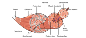
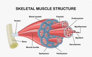
Skeletal muscle is composed of bundles of muscle fibers, each surrounded by connective tissue layers. This organization allows for efficient force generation and transmission.
Epimysium: Surrounds the entire muscle.
Perimysium: Surrounds each fascicle (bundle of muscle fibers).
Endomysium: Surrounds individual muscle fibers (cells).
Muscle Fiber (Cell): The basic contractile unit, containing myofibrils.
Myofibril: Cylindrical structures within muscle fibers, composed of repeating sarcomeres.
Sarcolemma: The cell membrane of a muscle fiber.
Tendon: Connects muscle to bone.
Blood Vessels & Capillaries: Supply nutrients and oxygen to muscle tissue.
Sarcomere Structure and Molecular Zones
Organization of the Sarcomere
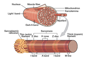
The sarcomere is the fundamental contractile unit of skeletal muscle, defined by specific molecular zones and boundaries.
Z-Discs (Z-Lines): Zig-zag boundaries at each end of a sarcomere; anchor thin actin filaments.
A-Band (Anisotropic): Dark band in the middle; width determined by thick myosin filaments (contains both myosin and overlapping actin).
I-Band (Isotropic): Light band spanning two adjacent sarcomeres; contains only thin actin filaments.
H-Zone: Lighter region in the center of the A-band; contains only thick myosin filaments (no actin overlap when muscle is resting).
M-Line: Dark line in the center of the H-zone; holds thick myosin filaments together.
Changes During Muscle Contraction
Muscle contraction involves the shortening of sarcomeres, resulting in movement. The following changes occur:
A-Band: Stays the same width (myosin filaments do not change size).
I-Band: Shortens/narrows (actin is pulled inward, decreasing the area where only actin exists).
H-Zone: Shortens and can disappear (actin moves into the center, overlapping myosin).
Z-Discs: Move closer together (overall sarcomere length decreases).
Mnemonic: "A-Band stays Average, but the H and I bands Hide."
Key Terms and Definitions
Sarcomere: The repeating contractile unit within a myofibril, defined by Z-discs.
Actin: Thin filament protein involved in muscle contraction.
Myosin: Thick filament protein responsible for force generation.
Sarcolemma: Muscle cell membrane.
Sarcoplasmic Reticulum: Organelle that stores and releases calcium ions for contraction.
Muscle Contraction Equation
The force generated by a muscle is proportional to the number of cross-bridges formed between actin and myosin:
F: Total muscle force
n: Number of cross-bridges
fcb: Force per cross-bridge
Example: Sarcomere Shortening
During contraction, the sarcomere shortens as actin filaments slide past myosin filaments, pulling Z-discs closer together and reducing the I-band and H-zone.
Additional info: The structural organization of muscle from whole muscle to sarcomere is essential for understanding muscle function and movement. The molecular zones of the sarcomere are critical for identifying changes during contraction and relaxation. The provided diagrams reinforce the hierarchical structure and molecular organization of skeletal muscle.
