 Indietro
Skeletal Muscle Physiology: Mechanisms of Muscle Contraction
Guida di studio - Note intelligenti
 Appunti personalizzati basati sui tuoi materiali, ampliati con definizioni chiave, esempi e contesto.
Skeletal Muscle Physiology
Overview of Muscle Contraction
Skeletal muscle contraction is a complex process involving the stimulation of muscle fibers by motor neurons, the transmission of electrical signals, and the interaction of contractile proteins. Understanding these mechanisms is essential for comprehending how voluntary movements are produced.
Muscle fibers are stimulated by motor neurons at specialized synapses called neuromuscular junctions.
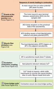
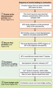
Contraction involves a sequence of events: excitation at the neuromuscular junction, muscle fiber excitation, excitation-contraction coupling, and the cross-bridge cycle.
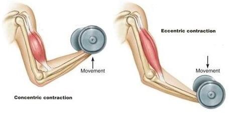
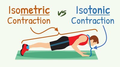
Muscle contractions can be classified as isometric or isotonic, with isotonic contractions further divided into concentric and eccentric types.
Sequence of Events Leading to Muscle Contraction
The process of muscle contraction can be divided into four main stages, each involving specific cellular and molecular events.
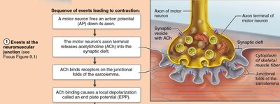
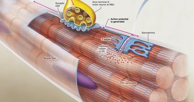
1. Events at the Neuromuscular Junction: The motor neuron releases acetylcholine (ACh), which binds to receptors on the muscle fiber, causing a local depolarization known as the end plate potential (EPP).
2. Muscle Fiber Excitation: The EPP triggers an action potential that travels across the sarcolemma (muscle cell membrane).
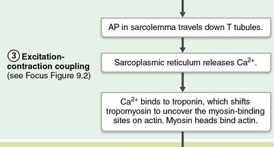
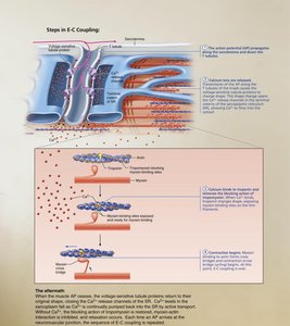
3. Excitation-Contraction Coupling: The action potential travels down T tubules, prompting the sarcoplasmic reticulum to release calcium ions (Ca2+), which bind to troponin and shift tropomyosin, exposing myosin-binding sites on actin.
4. Cross Bridge Cycle: Myosin heads bind to actin, leading to contraction via cross-bridge cycling.
Events at the Neuromuscular Junction
The neuromuscular junction is the site where a motor neuron communicates with a muscle fiber.
Acetylcholine (ACh) is released from the motor neuron into the synaptic cleft.
ACh binds to receptors on the sarcolemma, causing a local depolarization (EPP).
The effects of ACh are quickly terminated by the enzyme acetylcholinesterase, which breaks down ACh into acetic acid and choline.
Removal of ACh prevents continued muscle contraction without further nervous stimulation.
Muscle Fiber Excitation
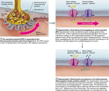
Muscle fiber excitation involves the generation and propagation of an action potential across the sarcolemma.
The EPP triggers an action potential that spreads across the sarcolemma.
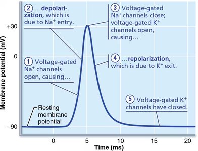
Voltage-gated sodium (Na+) channels open, causing depolarization.
Voltage-gated potassium (K+) channels open, causing repolarization.
Excitation-Contraction Coupling
Excitation-contraction coupling is the process by which an action potential leads to muscle contraction.
The action potential travels down T tubules, triggering the release of Ca2+ from the sarcoplasmic reticulum.
Ca2+ binds to troponin, causing tropomyosin to shift and expose myosin-binding sites on actin.
Myosin heads bind to actin, initiating contraction.
Cross Bridge Cycle
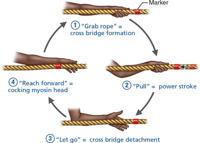
The cross bridge cycle describes the repetitive interactions between myosin and actin that result in muscle contraction.
Step 1: Cross Bridge Formation – Myosin heads bind to actin.
Step 2: Power Stroke – Myosin heads pivot, pulling actin filaments toward the center of the sarcomere.
Step 3: Cross Bridge Detachment – ATP binds to myosin, causing it to detach from actin.
Step 4: Cocking of Myosin Head – ATP is hydrolyzed, re-cocking the myosin head for another cycle.
Sarcomere Structure and Muscle Contraction
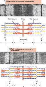
The sarcomere is the functional unit of skeletal muscle, composed of thick (myosin) and thin (actin) filaments.
During contraction, the sarcomere shortens as actin and myosin filaments slide past each other.
The distance between Z lines decreases, and the H zone and I band become narrower.
The Motor Unit
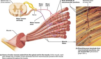
A motor unit consists of a motor neuron and all the muscle fibers it innervates.
When a motor neuron fires, all the muscle fibers in its motor unit contract simultaneously.
Motor units allow for graded control of muscle contraction strength.
Muscle Twitch and Graded Contractions
A muscle twitch is the response of a muscle to a single stimulus.
Graded contractions are produced by varying the frequency and strength of stimulation.
Muscle Tension and Load
Muscle tension is the force exerted by a contracting muscle.
Load is the opposing force exerted by the weight of the object to be moved.
Types of Muscle Contractions
Isotonic Contractions
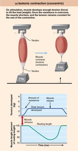
Isotonic contractions involve changes in muscle length while tension remains constant.
Concentric contraction: Muscle shortens and does work (e.g., lifting a weight).
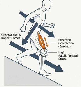
Eccentric contraction: Muscle lengthens while generating force (e.g., lowering a weight).
Isometric Contractions
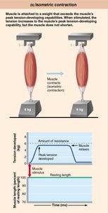
Isometric contractions occur when muscle tension increases but the muscle does not change length.
Occurs when the load is greater than the force the muscle can generate.
Important for maintaining posture and stabilizing joints.
Summary Table: Types of Muscle Contraction
Type | Muscle Length Change | Example |
|---|---|---|
Isotonic (Concentric) | Shortens | Lifting a weight |
Isotonic (Eccentric) | Lengthens | Lowering a weight, walking downhill |
Isometric | No change | Holding a weight steady, maintaining posture |
Key Terms and Definitions
Neuromuscular junction: Synapse between a motor neuron and a muscle fiber.
End plate potential (EPP): Local depolarization at the neuromuscular junction.
Excitation-contraction coupling: Process linking action potential to muscle contraction.
Cross bridge cycle: Repetitive interaction of myosin and actin during contraction.
Motor unit: Motor neuron and all muscle fibers it innervates.
Isotonic contraction: Muscle changes length while tension remains constant.
Isometric contraction: Muscle tension increases without length change.
Relevant Equations
Muscle tension: (Force equals mass times acceleration)
Membrane potential change:
Additional info:
Muscle soreness after eccentric contractions is due to microtrauma in muscle fibers.
Excitation-contraction coupling is essential for voluntary movement and is regulated by calcium ion dynamics.
