 Indietro
Skeletal Muscle Physiology: Membrane Potentials, Neuromuscular Junction, and Muscle Contraction
Guida di studio - Note intelligenti
 Appunti personalizzati basati sui tuoi materiali, ampliati con definizioni chiave, esempi e contesto.
Skeletal Muscle Physiology
Membrane Potentials
The membrane potential is a type of potential energy resulting from the separation of oppositely charged ions across the plasma membrane. This separation is essential for the function of excitable cells such as neurons and muscle fibers.
Ion concentration gradients: Differences in ion concentrations between the intracellular fluid (ICF) and extracellular fluid (ECF) create a charge separation.
Membrane ion permeability: The plasma membrane's selective permeability to ions, especially potassium (K+) and sodium (Na+), contributes to the membrane potential.
Measurement: Membrane potentials are measured in millivolts (mV).
Resting membrane potential typically ranges from -50 to -90 mV, indicating that the inside of the cell is negatively charged compared to the outside. This charge separation exists only at the plasma membrane.
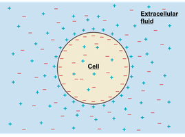
Why is the resting membrane potential negative? At rest, the membrane is more permeable to K+ than Na+ due to the abundance of K+ leak channels. K+ leaks out of the cell, making the inside more negative.
ICF vs. ECF: The concentration of K+ is higher inside the cell than outside, driving K+ out through leak channels.
Electrical Signals in Muscle Cells
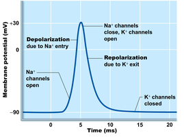
Electrical signals are changes in membrane potential away from the resting state, produced by excitable cells such as neurons and muscle fibers. These signals are generated by gated ion channels:
Chemically-gated ion channels
Voltage-gated ion channels
Mechanically-gated ion channels
Key terms:
Depolarization: Membrane potential becomes less negative.
Hyperpolarization: Membrane potential becomes more negative.
Repolarization: Membrane potential returns to resting value.
Neuromuscular Junction
Anatomy of the Neuromuscular Junction
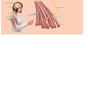
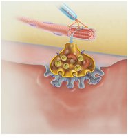
The neuromuscular junction is the region where a motor neuron contacts a skeletal muscle fiber, enabling voluntary muscle control.
Motor neuron: Contains a cell body, axon, and axon terminals.
Muscle fiber: Controlled by somatic motor neurons.
Communication Across the Neuromuscular Junction
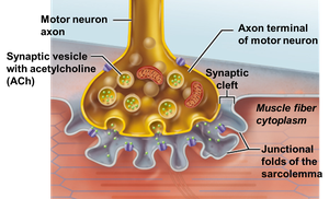
Communication at the neuromuscular junction involves the release of the neurotransmitter acetylcholine (ACh), which triggers muscle fiber excitation.
Action potential arrives at the axon terminal of the motor neuron.
Voltage-gated Ca2+ channels open, allowing Ca2+ to enter the axon terminal.
Ca2+ entry causes ACh to be released by exocytosis.
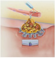
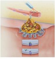
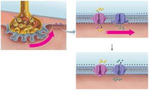
ACh diffuses across the synaptic cleft and binds to ACh receptors on the sarcolemma.
ACh binding opens chemically-gated ion channels, allowing Na+ to enter the muscle fiber, producing an end plate potential.
ACh effects are terminated by its breakdown via acetylcholinesterase.
Excitation-Contraction Coupling
Overview of Excitation-Contraction Coupling
Excitation-contraction coupling describes the events linking muscle fiber excitation (action potential) to contraction (cross bridge formation).
Action potential triggers a rise in cytosolic Ca2+ concentration.
Ca2+ directs cross bridge formation between actin and myosin.
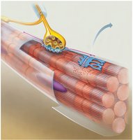
Steps in Excitation-Contraction Coupling
Action potential propagates along the sarcolemma and into T-tubules.
Voltage-sensitive tubule proteins change shape, opening Ca2+ release channels in the terminal cisterns.
Ca2+ flows into the cytosol, initiating contraction.
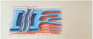
Role of Ca2+ in Muscle Contraction
Ca2+ binds to troponin, causing a conformational change that moves tropomyosin away from myosin binding sites on actin, allowing cross bridge formation.
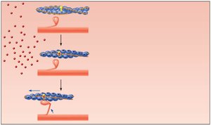
Cross Bridge Cycle
Steps of the Cross Bridge Cycle
The cross bridge cycle is responsible for sliding thin filaments (actin) along thick filaments (myosin), resulting in muscle contraction. The cycle is ATP-dependent and continues as long as ATP and Ca2+ are available.
Cross bridge formation: Energized myosin head attaches to actin.
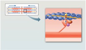
Power stroke: ADP and Pi are released; myosin head pivots, pulling actin toward the M line.
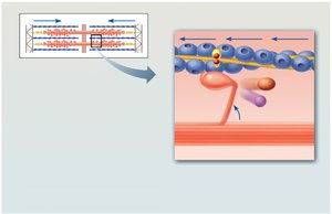
Cross bridge detachment: ATP binds to myosin, causing detachment from actin.
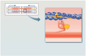
Myosin head re-energization: ATP is hydrolyzed to ADP and Pi, re-cocking the myosin head.
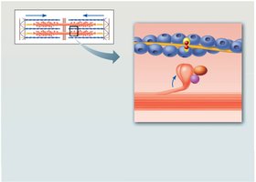
The cycle continues as long as ATP and Ca2+ are present. If ATP is unavailable, the cycle stops between steps 2 and 3.
Ending the Cross Bridge Cycle
Ca2+ pumps in the sarcoplasmic reticulum use ATP to transport Ca2+ back into terminal cisterns.
As cytosolic Ca2+ falls, troponin loses Ca2+ and tropomyosin blocks myosin binding sites, ending contraction.
Cross Bridge Cycle and Rigor Mortis
After death, ATP levels decline, causing Ca2+ to leak out and promote cross bridge formation. Without ATP, cross bridges cannot detach, resulting in muscle stiffening known as rigor mortis.
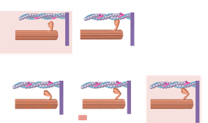
Summary Table: Steps of the Cross Bridge Cycle
Step | Description |
|---|---|
1. Cross bridge formation | Myosin head binds to actin |
2. Power stroke | Myosin head pivots, pulling actin |
3. Cross bridge detachment | ATP binds to myosin, causing detachment |
4. Myosin head re-energization | ATP hydrolysis re-cocks myosin head |
Key Equations
Nernst Equation: Used to calculate equilibrium potential for an ion.
Membrane Potential:
Review Questions
What are the two requirements needed to establish a membrane potential?
Why are resting membrane potentials negatively charged?
What types of ion channels are responsible for producing electrical signals?
Explain how communication across the neuromuscular junction occurs.
What is excitation-contraction coupling?
How does an action potential cause the terminal cisterns to release Ca2+ into the cytosol?
How does a rise in cytosolic Ca2+ lead to the binding of myosin heads to actin?
What are the four steps of the cross bridge cycle?
How does the return of Ca2+ to the terminal cisterns cause a muscle to relax?
What causes rigor mortis to develop after death?
Additional info: Academic context was added to clarify the mechanisms of membrane potential, excitation-contraction coupling, and rigor mortis, as well as to provide relevant equations and a summary table for the cross bridge cycle.
