 Indietro
Skeletal Muscle Tissue Structure and Function: Study Notes
Guida di studio - Note intelligenti
 Appunti personalizzati basati sui tuoi materiali, ampliati con definizioni chiave, esempi e contesto.
Skeletal Muscle Tissue: Structure and Function
Overview of Skeletal Muscle Tissue
Skeletal muscle is a specialized tissue responsible for voluntary movements, posture, and heat generation. It is composed of elongated, multinucleate, striated cells called muscle fibers, and is organized into organs that include blood vessels, nerve fibers, and connective tissue sheaths.
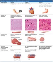
Location: Attached to and covering the skeleton
Control: Voluntary (conscious control)
Cell Structure: Long, cylindrical, multinucleate, striated
Functions of Skeletal Muscle
Skeletal muscle performs several essential functions in the human body:
Produce movement: Responsible for locomotion and manipulation of the environment.
Maintain posture and body position: Continuous muscle contractions stabilize the body.
Stabilize joints: Muscle tone reinforces and stabilizes joints.
Generate heat: Muscle activity produces heat, helping maintain body temperature.
Connective Tissue Sheaths of Skeletal Muscle
Three Connective Tissue Sheaths
Each skeletal muscle and its fibers are covered by connective tissue sheaths that support and protect the muscle:
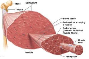
Epimysium: Dense irregular connective tissue surrounding the entire muscle.
Perimysium: Fibrous connective tissue surrounding groups of muscle fibers called fascicles.
Endomysium: Fine areolar connective tissue surrounding each individual muscle fiber.
Muscle Attachments
Muscles attach to bones in two main ways:
Direct attachment: The epimysium fuses directly with the periosteum of a bone. This is less common.
Indirect attachment: Connective tissue extends beyond the muscle as a tendon (rope-like) or aponeurosis (sheet-like), which then attaches to the bone's periosteum.
Microscopic Anatomy of Skeletal Muscle Fibers
Structure of Muscle Fibers
Muscle fibers are the basic cellular units of skeletal muscle. They contain specialized structures for contraction and energy storage:
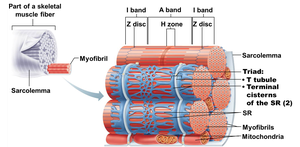
Sarcolemma: The plasma membrane of a muscle fiber.
Sarcoplasm: The cytoplasm of a muscle fiber, containing organelles, glycosomes (glycogen storage), and myoglobin (oxygen-binding protein).
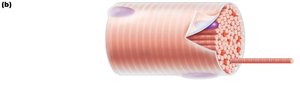
Myofibrils: Long, rod-like structures that fill most of the muscle fiber's volume and are responsible for contraction.
Sarcoplasmic reticulum (SR): Specialized smooth endoplasmic reticulum that stores and releases calcium ions (Ca2+).
Myofibrils and Sarcomeres
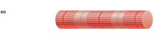
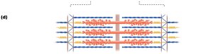
Myofibrils are composed of repeating units called sarcomeres, which are the functional contractile units of muscle fibers. The arrangement of myofilaments within sarcomeres gives skeletal muscle its striated appearance.
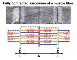
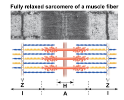
Sarcomere: Extends from one Z disc to the next; contains thick and thin filaments.
A band: Dark region; length of thick filaments.
I band: Light region; contains only thin filaments.
H zone: Lighter region in the middle of the A band; contains thick filaments not overlapped by thin filaments.
M line: Center of the H zone; holds thick filaments together.
Z disc: Boundary of each sarcomere; anchors thin filaments.
Proteins of the Sarcomere
The sarcomere contains several key proteins:
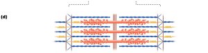
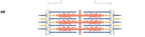
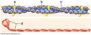
Thick filaments: Composed of myosin molecules, each with a tail and two heads. The heads have binding sites for actin and ATP.
Thin filaments: Composed of actin (with myosin-binding sites), tropomyosin (blocks binding sites in relaxed muscle), and troponin (regulates tropomyosin position).
Titin: A large protein that anchors thick filaments to the Z disc and provides elasticity.
Thick and Thin Filament Structure
Myosin: Each molecule has two heavy chains (forming the tail and heads) and four light chains. The heads bind to actin and ATP, enabling contraction.
Actin: Forms a double helix; each monomer has a myosin-binding site.
Tropomyosin: Covers actin's myosin-binding sites in relaxed muscle.
Troponin: Binds Ca2+ and moves tropomyosin away from binding sites during contraction.
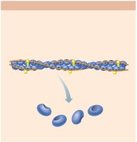
Sliding Filament Model of Contraction
Mechanism of Muscle Contraction
The sliding filament model explains how muscles contract at the molecular level:
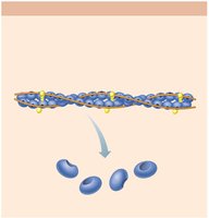
Myosin heads bind to actin, forming cross bridges.
Cross bridges rotate, pulling thin filaments toward the center of the sarcomere.
Thin filaments slide past thick filaments, shortening the sarcomere and thus the muscle fiber.
During contraction, the I bands and H zone shorten, the A band remains the same length, and the Z discs move closer together.
Sarcoplasmic Reticulum and T Tubules
Sarcoplasmic Reticulum (SR)
The SR is a specialized smooth endoplasmic reticulum that regulates intracellular Ca2+ levels, which are essential for muscle contraction.
SR forms a network around each myofibril.
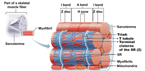
Terminal cisterns are enlarged areas of the SR that store Ca2+.
T Tubules and Triads
T tubules are invaginations of the sarcolemma that penetrate into the muscle fiber, allowing action potentials to quickly reach the interior. A triad consists of a T tubule flanked by two terminal cisterns of the SR, facilitating the coupling of excitation and contraction.
Review Table: Key Proteins of the Thin Filament
Label | Protein | Function |
|---|---|---|
a | Actin | Main component of thin filament; contains myosin-binding sites |
b | Tropomyosin | Covers myosin-binding sites on actin in relaxed muscle |
c | Troponin | Binds Ca2+ and moves tropomyosin to expose binding sites |
d | Myosin | Main component of thick filament; binds to actin and ATP |
Summary Table: Changes During Muscle Contraction
Band/Zone | Change During Contraction |
|---|---|
I band | Shortens |
A band | Remains the same length |
H zone | Disappears |
Z discs | Move closer together |
Degree of overlap | Increases |
Filament length | Does not change |
Key Equations and Concepts
ATP hydrolysis: Provides energy for myosin head movement
Calcium release: Triggered by action potentials, Ca2+ binds to troponin, initiating contraction.
Additional Info
Muscle triad: The triad structure (T tubule + 2 terminal cisterns) is essential for rapid and coordinated muscle contraction.
Ca2+ storage: In a relaxed muscle fiber, Ca2+ is stored in the sarcoplasmic reticulum.
