 Indietro
Skeletal Muscle Tissue: Structure, Function, and Contraction Mechanisms
Guida di studio - Note intelligenti
 Appunti personalizzati basati sui tuoi materiali, ampliati con definizioni chiave, esempi e contesto.
Skeletal Muscle Tissue
General Function and Anatomical Organization
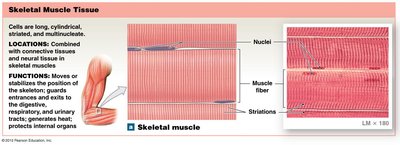
Skeletal muscle tissue is specialized for contraction, enabling movement, maintaining posture, regulating body temperature, and guarding entrances and exits of the body. It is composed of long, cylindrical, multinucleated cells and is organized hierarchically from the organ level down to the molecular level.
Main Functions: Movement, posture, heat production, sphincter control, soft tissue support, and nutrient storage.
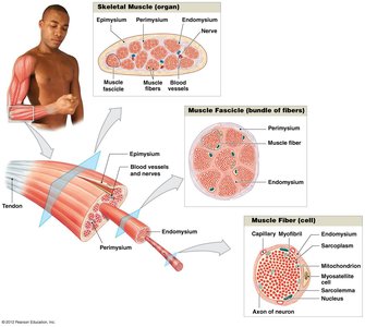
Organization: Muscle (organ) → Fascicle (tissue) → Muscle fiber (cell) → Myofibril (organelle) → Sarcomere (protein complex) → Actin & myosin (proteins).
Types of Muscle Tissue
There are three main types of muscle tissue in the human body, each with distinct structural and functional characteristics.
Skeletal Muscle: Voluntary, striated, multinucleated; attached to bones for movement.
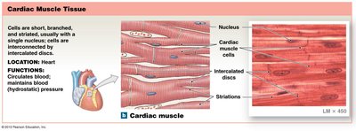
Cardiac Muscle: Involuntary, striated, branched; found only in the heart.
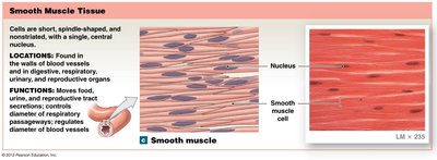
Smooth Muscle: Involuntary, non-striated, spindle-shaped; found in walls of hollow organs.
Development and Structure of Skeletal Muscle Fibers
Development of Muscle Fibers
Skeletal muscle fibers develop from the fusion of mesodermal cells called myoblasts. Mature muscle fibers are multinucleated and can reach up to 30 cm in length.
Myoblasts: Embryonic cells that fuse to form muscle fibers.
Myosatellite cells: Aid in muscle repair and regeneration.
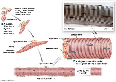
Structure of a Skeletal Muscle Fiber
Each muscle fiber contains multiple nuclei, a sarcolemma (cell membrane), and numerous myofibrils, which are composed of repeating units called sarcomeres.
Myofibrils: Bundles of protein filaments responsible for contraction.
Sarcolemma: Plasma membrane of the muscle fiber.
Sarcoplasm: Cytoplasm of the muscle fiber.
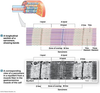
Sarcomere Structure and Muscle Contraction
Anatomy of the Sarcomere
The sarcomere is the fundamental contractile unit of skeletal muscle, defined by the region between two Z lines. It contains thick (myosin) and thin (actin) filaments arranged in a precise pattern.
Z line: Boundary of the sarcomere; anchors thin filaments.
M line: Center of the sarcomere; anchors thick filaments.
A band: Region containing thick filaments.
I band: Region containing only thin filaments.
H zone: Region containing only thick filaments.
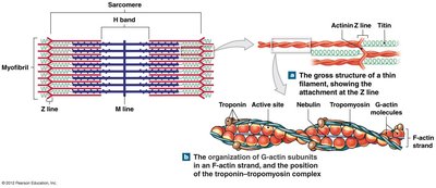
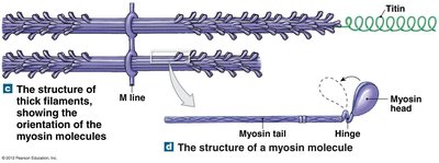
Changes During Muscle Contraction
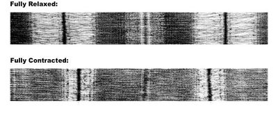
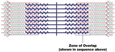
Muscle contraction involves the sliding of thin filaments past thick filaments, shortening the sarcomere. The H band and I band decrease in length, while the A band remains unchanged.
Shortening: H band and I band become shorter; Z lines move closer together.
Constant: A band and filament lengths remain the same.
Molecular Mechanisms of Muscle Contraction
Neuromuscular Junction and Signal Transmission
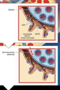
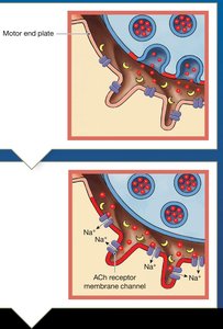
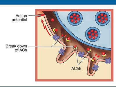
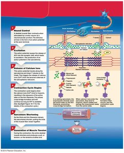
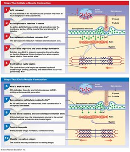
The neuromuscular junction is where a motor neuron communicates with a muscle fiber, initiating contraction via the release of acetylcholine (ACh).
ACh Release: Neurotransmitter released from synaptic vesicles.
Action Potential: Electrical signal travels along the sarcolemma and T tubules.
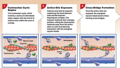
Calcium Release: Sarcoplasmic reticulum releases Ca2+ ions, triggering contraction.
Contraction Cycle
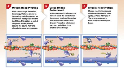
The contraction cycle consists of several steps, including cross-bridge formation, power stroke, and detachment, all requiring ATP.
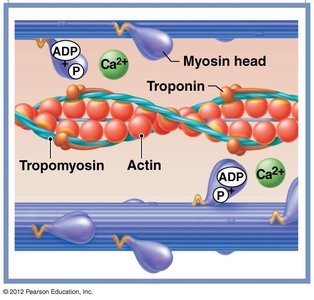
Cross-Bridge Formation: Myosin heads bind to actin.
Power Stroke: Myosin head pivots, pulling actin filament.
Detachment: ATP binds to myosin, causing release from actin.
Reactivation: ATP hydrolysis re-cocks the myosin head.
Energy Requirements
ATP is essential for both muscle contraction and relaxation. The hydrolysis of ATP provides energy for the power stroke and for detaching myosin from actin.
ATP Hydrolysis:
Relaxation: ATP is required to reset the myosin head and pump Ca2+ back into the sarcoplasmic reticulum.
Analogy: Sarcomere as a Mousetrap
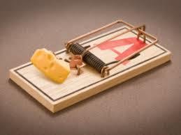
The sarcomere can be compared to a mousetrap, where the myosin head acts as the lever, actin as the platform, tropomyosin as the bait, and ATP as the mechanism to reset the trap.
Myosin head: Lever
Actin: Platform
Tropomyosin: Bait
ATP: Resets/releases lever
Pathology: Duchenne Muscular Dystrophy (DMD)
Overview and Clinical Features
Duchenne Muscular Dystrophy is a severe, progressive muscle-wasting disease caused by the absence of dystrophin, a protein that stabilizes muscle fibers during contraction. It is inherited in an X-linked recessive manner, primarily affecting males.
Symptoms: Progressive muscle weakness, loss of ambulation by age 12, cardiac and respiratory complications.
Pathophysiology: Lack of dystrophin leads to muscle fiber damage and degeneration.
Life Expectancy: Median 20–30 years; most common causes of death are cardiac or respiratory failure.
Summary Table: Muscle Tissue Types
Type | Structure | Location | Function |
|---|---|---|---|
Skeletal | Long, cylindrical, multinucleated, striated | Attached to bones | Movement, posture, heat production |
Cardiac | Short, branched, striated, single nucleus | Heart | Circulates blood, maintains pressure |
Smooth | Spindle-shaped, non-striated, single nucleus | Walls of hollow organs | Moves food, regulates vessel diameter |
Additional info:
Muscle contraction is regulated by the interaction of actin, myosin, troponin, and tropomyosin, with calcium ions playing a critical role in exposing binding sites.
ATP is required for both contraction and relaxation, emphasizing the importance of energy metabolism in muscle physiology.
