 Indietro
Skeletal Muscle Tissue: Structure, Function, and Physiology
Guida di studio - Note intelligenti
 Appunti personalizzati basati sui tuoi materiali, ampliati con definizioni chiave, esempi e contesto.
Skeletal Muscle Tissue
Types of Muscle Tissue
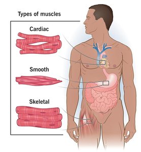
Muscle tissue is specialized for contraction and is essential for movement and various bodily functions. There are three main types of muscle tissue:
Skeletal muscle: Voluntary control, attached to bones, responsible for movement.
Cardiac muscle: Involuntary, found only in the heart.
Smooth muscle: Involuntary, located in the walls of hollow organs.
Functions of Skeletal Muscle Tissue
Skeletal muscle tissue performs several critical functions in the body:
Produce body movement: Muscle tendons pull and move bones.
Maintain posture and body position: Stabilizes joints.
Support soft tissue: Surrounds, supports, and shields internal structures.
Guard body entrances/exits: Sphincters provide voluntary control of swallowing, defecation, and urination.
Maintain body temperature: Contraction generates heat.
Store nutrients: Muscle proteins can break down to release amino acids for energy or glucose synthesis.
Structure of Skeletal Muscle
Organizational Levels
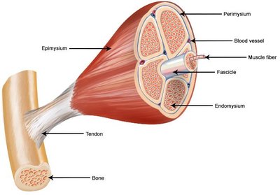
Skeletal muscle is an organ composed of muscle tissue, connective tissue, nerves, and blood vessels. The structure is organized into several layers:
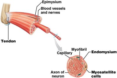
Epimysium: Dense sheath of collagen fibers surrounding the entire muscle, separating it from other tissues.
Perimysium: Fibrous layer dividing muscle into compartments called fascicles; contains blood vessels and nerves.
Endomysium: Thin layer of areolar connective tissue around each muscle fiber; contains capillaries, nerves, and myosatellite cells (stem cells for repair).
Muscle fascicle: Bundle of muscle fibers.
Muscle fiber: Individual muscle cell containing myofibrils.
Skeletal Muscle Development
Skeletal muscle fibers develop from embryonic cells called myoblasts:
Myoblasts fuse to form multinucleate cells that differentiate into muscle fibers.
Some myoblasts remain as myosatellite cells in the endomysium, aiding in muscle repair.
Mature muscle fibers are large, with diameters up to 100 μm and lengths up to 30 cm.
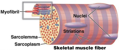
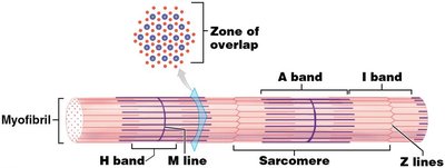
Skeletal Muscle Fiber Structure
Myofibrils and Myofilaments
Each muscle fiber contains numerous myofibrils, which are bundles of protein filaments:
Myofibrils: Cylindrical structures running the length of the muscle fiber; responsible for striations (alternating light and dark bands).
Myofilaments: Protein filaments within myofibrils; two main types:
Thin filaments: Mostly composed of actin.
Thick filaments: Mostly composed of myosin.
Sarcomeres and Striations
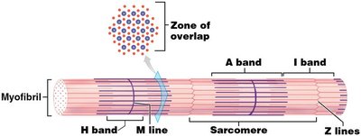
The functional unit of a muscle fiber is the sarcomere:
Each myofibril contains about 10,000 sarcomeres.
Sarcomeres are ~2 μm at resting length.
Striations are due to the arrangement of thick and thin filaments.
Key regions:
Z lines: Junction of adjacent sarcomeres; connect thin filaments.
I band: Lighter band with only thin filaments.
A band: Dark region with thick filaments.
Zone of overlap: Area where thick and thin filaments overlap.
M line: Center of A band; connects thick filaments.
H band: Lighter region on each side of M line with only thick filaments.
Sarcolemma and T Tubules
The sarcolemma is the plasma membrane of a muscle fiber, and T tubules are extensions that help transmit action potentials:
Sarcolemma is selectively permeable, allowing uneven distribution of charges.
T tubules are continuous with the sarcolemma and encircle the sarcomere, facilitating rapid transmission of electrical signals.
Sarcoplasmic Reticulum (SR)
The SR is similar to the smooth endoplasmic reticulum and stores calcium ions:
Terminal cisternae are enlarged sections adjacent to T tubules.
Triad = pair of terminal cisternae and one T tubule.
SR releases calcium ions to initiate muscle contraction.
Sliding Filament Theory
Structure of Thin and Thick Filaments
Thin filaments (Actin): Composed of F-actin (double strand of G-actin), nebulin, tropomyosin (blocks myosin binding sites), and troponin (binds calcium).
Thick filaments (Myosin): Composed of ~300 myosin molecules, titin (connects thick filaments to Z lines), and myosin heads that form cross-bridges with actin.
Sliding Filament Theory of Muscle Contraction
Muscle contraction occurs when thin filaments slide over thick filaments:
H and I bands get smaller; zones of overlap get larger.
Z lines move closer together; A band width remains unchanged.
All sarcomeres in each myofibril contract simultaneously, shortening the muscle fiber.
Excitable Plasma Membrane and Action Potential
Membrane Potentials
All cells are polarized, with unequal distribution of positive and negative charges across the plasma membrane.
Inside of cell is slightly negative compared to outside.
Membrane potential is measured in millivolts (mV).
Movement of Sodium and Potassium
Leak channels allow slow flow of Na+ and K+ down their concentration gradients.
Sodium–potassium ion pumps export 3 Na+ and import 2 K+.
Action Potential
Small increase in membrane permeability to Na+ moves membrane potential to threshold (-55 mV).
Voltage-gated channels open; Na+ rushes in (depolarization).
Membrane potential reaches +30 mV; Na+ channels close, K+ channels open (repolarization).
Repolarization returns to resting membrane potential.
Na+/K+ pump restores original distribution.
Neuromuscular Junction (NMJ)
NMJ Components
Axon terminal of motor neuron contains vesicles with acetylcholine (ACh).
Motor end plate of muscle fiber has junctional folds with ACh receptors.
Synaptic cleft is the space between axon terminal and motor end plate.
Acetylcholinesterase (AChE) breaks down ACh.
Events at Neuromuscular Junction
Action potential arrives at axon terminal; ACh released.
ACh diffuses across synaptic cleft, binds to receptors, changes Na+ permeability.
Na+ influx generates action potential in sarcolemma; ACh is broken down.
Action potential spreads across sarcolemma.
Action potential moves down T tubules, changes SR permeability.
SR releases Ca2+ into sarcomere, initiating contraction (excitation-contraction coupling).
Skeletal Muscle Fiber Contraction
Contraction Cycle
Resting sarcomere: Myosin heads are cocked, ready to attach to actin (uses ATP).
Contraction cycle begins: Ca2+ arrives from SR.
Active sites exposed: Ca2+ binds to troponin, moves tropomyosin, exposes actin sites.
Cross-bridges form: Myosin heads bind to actin.
Myosin heads pivot: Power stroke toward M line.
Cross-bridges detach: New ATP binds, myosin releases from actin.
Myosin reactivates: ATP split, energy used to recock head.
Cycle repeats while Ca2+ and ATP are available; when stimulus ends, Ca2+ returns to SR, active sites covered.
Excitation-Contraction Coupling
The link between generation of action potential in sarcolemma and start of muscle contraction occurs in six steps:
Neural control: Action potential in motor neuron starts process at NMJ.
Excitation: ACh release leads to action potential in sarcolemma.
Calcium ion release: Action potential triggers Ca2+ release from SR.
Contraction cycle begins: Ca2+ binds troponin, exposes actin sites, cross-bridges form.
Sarcomeres shorten: Sliding filaments shorten sarcomeres.
Muscle tension produced: Muscle fiber shortening causes muscle contraction.
Functional Properties of Skeletal Muscle
Sarcomere Length–Tension Relationship
Tension is greatest when muscle fibers are stimulated at optimal length:
Maximum tension occurs when actin and myosin overlap optimally.
If sarcomere is too short, filaments overlap too much; if too long, not enough overlap.
Muscle and connective tissues maintain sarcomere length within a safe range (75–130% of optimal).
Muscle Twitch
A muscle twitch is a single stimulus-contraction-relaxation cycle in a muscle fiber, shown on a myogram:
Latent period: Action potential stimulates sarcolemma; Ca2+ released; no tension yet.
Contraction period: Ca2+ binds troponin; cross-bridge cycling; tension develops to peak.
Relaxation phase: Ca2+ drops; cross-bridges detach; tension returns to resting levels.
Muscle Tension
Tension produced depends on amount of tension per fiber and number of fibers stimulated.
Patterns of muscle tension:
Treppe: Increasing maximum tension with repeated stimulation after relaxation.
Wave summation: Addition of one twitch to another before relaxation ends.
Incomplete tetanus: Rapid contraction/relaxation cycles, near-peak tension.
Complete tetanus: No relaxation phase, peak tension, continuous contraction.
Motor Units and Recruitment
Motor unit: Single motor neuron and all muscle fibers it controls.
Activation of more motor units increases tension; smaller units activated first, then larger ones.
Isotonic and Isometric Contractions
Isotonic contraction: Tension rises, muscle length changes (e.g., lifting, walking).
Concentric contraction: Muscle shortens, tension exceeds load.
Eccentric contraction: Muscle lengthens, peak tension less than load.
Isometric contraction: Muscle length does not change, tension never exceeds load (e.g., postural muscles).
Energy for Muscle Contraction
Sources of ATP
Glycolysis: Anaerobic breakdown of glucose to pyruvate; produces 2 ATP per glucose.
Aerobic metabolism: Occurs in mitochondria; produces 15 ATP per pyruvate; supplies 95% of ATP in resting cells.
Muscle Metabolism
At rest: Low ATP demand; mitochondria produce surplus ATP; fatty acids and glucose used to make creatine phosphate and glycogen.
Moderate activity: ATP demand increases; relies on aerobic metabolism; no fatigue until reserves exhausted.
Peak activity: High ATP demand; mitochondria provide ~1/3 ATP; rest from glycolysis; excess pyruvate converts to lactate, causing fatigue.
Muscle Fatigue and Recovery
Fatigue: Muscle can no longer perform at required level; major factor is decline in pH.
Recovery period: Time needed to return muscle fibers to preexertion levels.
Oxygen debt (EPOC): Amount of oxygen required to restore normal conditions; used to restore ATP, creatine phosphate, glycogen, and convert lactate to glucose.
Skeletal Muscle Fiber Types
Classification of Muscle Fibers
Fast fibers: Large diameter, densely packed myofibrils, large glycogen reserves, few mitochondria; powerful, fatigue rapidly.
Slow fibers: Smaller diameter, longer sustained contractions, mostly aerobic ATP production, extensive capillary network, dark red appearance.
Intermediate fibers: Resemble fast fibers, little myoglobin, pale, more capillaries, more fatigue-resistant than fast fibers.
Fiber Type | Diameter | ATP Production | Fatigue Resistance | Color |
|---|---|---|---|---|
Fast | Large | Anaerobic | Low | Pale |
Slow | Small | Aerobic | High | Dark Red |
Intermediate | Medium | Mixed | Moderate | Pale |
Example: Fast fibers are used for quick, powerful movements (e.g., sprinting), while slow fibers are used for endurance activities (e.g., marathon running). Additional info: Table content inferred for clarity and completeness.
