 Indietro
Skeletal System: Structure, Function, and Anatomy
Guida di studio - Note intelligenti
 Appunti personalizzati basati sui tuoi materiali, ampliati con definizioni chiave, esempi e contesto.
Skeletal System Overview
Mobility and Components
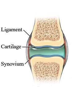
The skeletal system is essential for mobility, providing the framework for purposeful physical movement. Optimal mobility depends on bones, joints, articular cartilage, tendons, ligaments, and skeletal muscle. Changes in mobility can impact overall health, and healthcare professionals play a key role in promoting mobility and preventing complications from immobility.
Bones: Provide structure and support.
Joints: Allow movement between bones.
Cartilages: Cushion and support joints.
Ligaments: Connect bones and stabilize joints.
Functions of the Skeletal System
Major Functions
The skeletal system performs several critical functions:
Support: Provides the structural framework for the body.
Protection: Shields vital organs (e.g., skull protects the brain, ribs protect the heart and lungs).
Movement: Acts as anchorage for muscles, enabling movement.
Storage: Stores minerals (calcium, phosphate), growth factors, and triglycerides.
Hormone Production: Osteocalcin regulates bone formation and insulin secretion.
Blood Cell Formation (Hematopoiesis): Occurs in bone marrow.
Organization of the Skeleton
Axial and Appendicular Skeleton
The human skeleton is divided into two main groups:
Axial Skeleton: Includes the skull, vertebral column, and thoracic cage.
Appendicular Skeleton: Includes the pectoral girdle, upper limbs, pelvic girdle, and lower limbs.
Axial Skeleton: Skull
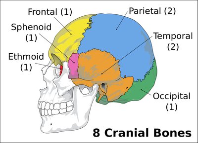
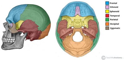
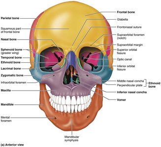
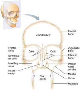
The skull consists of 22 bones, divided into cranial and facial bones. Cranial bones protect the brain, while facial bones form the structure of the face.
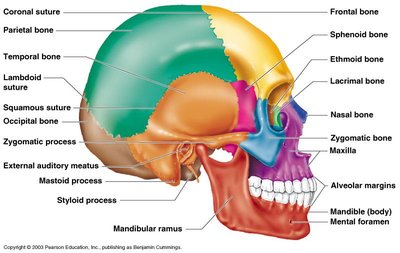
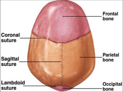
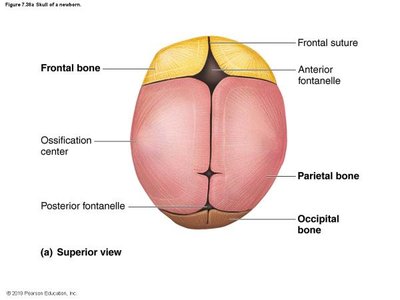
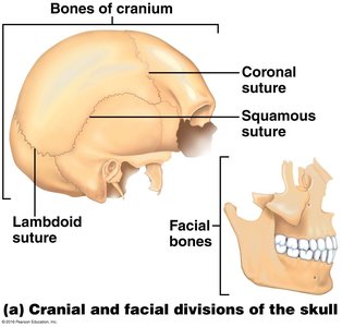
Axial Skeleton: Hyoid Bone
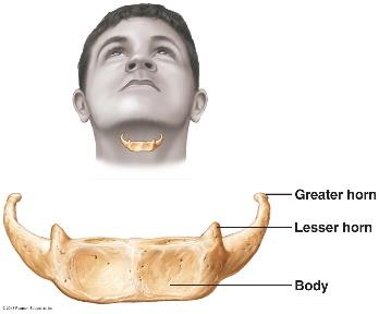
The hyoid bone is unique, located in the neck and not directly attached to other bones. It supports the tongue and is involved in swallowing.
Axial Skeleton: Thoracic Cage
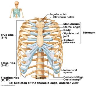
The thoracic cage consists of ribs, sternum, and thoracic vertebrae. It protects the heart and lungs and supports the upper body.
Axial Skeleton: Vertebral Column
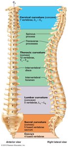
The vertebral column is composed of cervical, thoracic, lumbar, sacral, and coccygeal regions. It supports the body and protects the spinal cord.
Appendicular Skeleton
The appendicular skeleton includes the bones of the limbs and girdles that attach them to the axial skeleton.
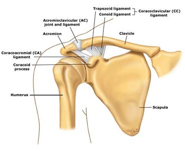
Pectoral Girdle: Clavicle and scapula.
Upper Limbs: Humerus, radius, ulna, carpals, metacarpals, phalanges.
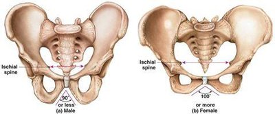
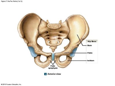
Pelvic Girdle: Hip bones (ilium, ischium, pubis).
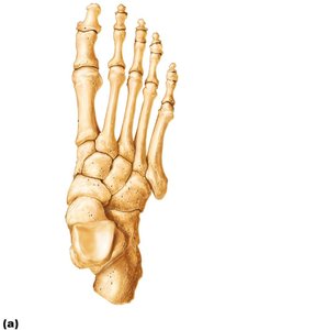
Lower Limbs: Femur, tibia, fibula, tarsals, metatarsals, phalanges.
Classification of Bones by Shape
Bone Types
Long Bones: Longer than they are wide (e.g., femur, humerus).
Short Bones: Cube-shaped (e.g., carpals, tarsals).
Flat Bones: Thin, flattened, and usually curved (e.g., sternum, skull).
Irregular Bones: Complex shapes (e.g., vertebrae, hip bones).
Bone Structure and Anatomy
Gross Anatomy
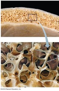
Bones have two main textures:
Compact Bone: Dense outer layer, smooth and solid.
Spongy Bone (Trabecular Bone): Honeycomb structure with trabeculae.
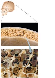
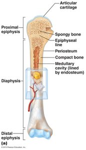
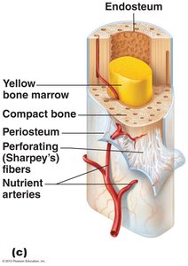
Microscopic Anatomy
Bones contain several cell types:
Osteogenic Cells: Stem cells in bone membranes.
Osteoblasts: Bone-forming cells.
Osteocytes: Mature bone cells that maintain bone matrix.
Bone Lining Cells: Flat cells on bone surfaces.
Osteoclasts: Bone-resorbing cells.
Compact Bone Structure
The structural unit of compact bone is the osteon (Haversian system), consisting of concentric lamellae surrounding a central canal.
Spongy Bone Structure
Spongy bone contains trabeculae aligned along lines of stress, with capillaries in the endosteum supplying nutrients.
Bone Development and Growth
Ossification (Osteogenesis)
Bone tissue formation occurs through two processes:
Endochondral Ossification: Bone forms by replacing hyaline cartilage (most bones).
Intramembranous Ossification: Bone develops from fibrous membrane (cranial bones, clavicles).
Postnatal Bone Growth
Interstitial Growth: Increases length of long bones via epiphyseal plate.
Appositional Growth: Increases thickness of bones throughout life.
Hormonal Regulation
Growth Hormone: Stimulates bone growth in childhood.
Thyroid Hormone: Modulates growth hormone activity.
Sex Hormones: Promote growth spurts and epiphyseal plate closure at puberty.
Bone Homeostasis and Remodeling
Remodeling and Calcium Homeostasis
Bone remodeling involves continuous bone resorption and deposition, regulated by calcium homeostasis and mechanical stress.
Osteoclasts: Break down bone, releasing calcium into blood.
Osteoblasts: Deposit new bone matrix.
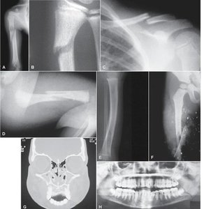
Bone Injury and Repair
Fractures and Healing
Fractures are breaks or cracks in bones, classified as closed/simple or open/compound. Bone repair occurs in four steps:
Hematoma Formation: Blood-filled swelling at fracture site.
Fibrocartilage Callus Formation: New capillaries and collagen fibers stabilize the fracture.
Bony Callus Formation: Spongy bone replaces fibrocartilage callus.
Bone Remodeling: Bone is remodeled to restore strength and shape.
Cartilage: Structure, Types, and Locations
Types of Cartilage
Hyaline Cartilage: Provides support, flexibility, and resilience; most abundant.
Elastic Cartilage: Contains elastic fibers; found in external ear and epiglottis.
Fibrocartilage: Thick collagen fibers; great tensile strength; found in intervertebral discs and menisci.
Summary Table: Bone Cell Types
Cell Type | Function |
|---|---|
Osteogenic Cells | Stem cells; differentiate into osteoblasts |
Osteoblasts | Bone-forming cells; secrete osteoid |
Osteocytes | Mature bone cells; maintain matrix |
Bone Lining Cells | Maintain bone matrix on surfaces |
Osteoclasts | Bone-resorbing cells; break down bone |
Summary Table: Bone Classification by Shape
Type | Example |
|---|---|
Long Bone | Femur, Humerus |
Short Bone | Carpals, Tarsals |
Flat Bone | Sternum, Skull |
Irregular Bone | Vertebrae, Hip Bones |
Key Equations
Calcium Homeostasis
Blood calcium levels are maintained within a narrow range:
Additional info:
Bone remodeling is influenced by mechanical stress and calcium/vitamin D levels.
Blood homeostasis is maintained at the cost of bone structure if necessary.
