 Indietro
Skeletal System: Structure, Function, and Development
Guida di studio - Note intelligenti
 Appunti personalizzati basati sui tuoi materiali, ampliati con definizioni chiave, esempi e contesto.
Skeletal System Overview
Structure and Function of the Skeletal System
The skeletal system provides the framework for the human body, supporting soft tissues, protecting vital organs, and facilitating movement. It also serves as a reservoir for minerals and houses sites for blood cell formation.
Support: Bones support the body and soft organs.
Protection: Protects the brain, spinal cord, and vital organs.
Movement: Bones act as levers for muscle action.
Mineral Storage: Stores calcium, phosphorus, and growth factors.
Blood Cell Formation: Hematopoiesis occurs in red bone marrow.
Triglyceride Storage: Fat is stored in bone cavities.
Hormone Production: Osteocalcin regulates insulin secretion and energy expenditure.
Key Terms Related to the Skeletal System
Cartilage and Associated Structures
The human skeleton initially consists of cartilage, which is gradually replaced by bone except in areas requiring flexibility. Cartilage is avascular and lacks nerves, and is surrounded by the perichondrium, a dense connective tissue layer that supplies nutrients.
Chondrocytes: Cartilage cells encased in lacunae within a jelly-like matrix.
Perichondrium: Surrounds cartilage, resists expansion, and supplies nutrients.
Types of Skeletal Cartilage
Three Types of Cartilage
Cartilage types differ in composition and location, each serving specific functions in the skeleton.
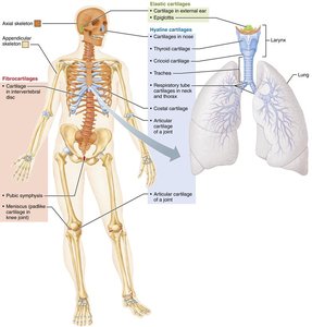
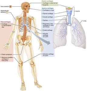
Hyaline Cartilage: Provides support, flexibility, and resilience; most abundant; contains only collagen fibers. Found in articular (joints), costal (ribs), respiratory (larynx, trachea), and nasal cartilage.
Elastic Cartilage: Similar to hyaline but contains elastic fibers; found in the external ear and epiglottis.
Fibrocartilage: Contains thick collagen fibers for tensile strength; found in menisci of knee, pubic symphysis, and intervertebral discs.
Classification of Bones
Bone Classification by Location and Shape
Bones are classified based on their location in the skeleton and their shape. The human skeleton has 206 named bones, divided into axial and appendicular groups.
Axial Skeleton: Long axis of the body; includes skull, vertebral column, and rib cage.
Appendicular Skeleton: Bones of limbs and girdles attaching limbs to the axial skeleton.
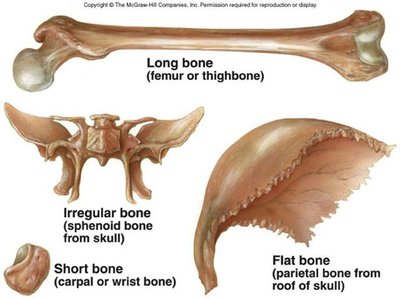
Four Types of Bone Shapes
Long Bones: Longer than wide, with a medullary cavity; e.g., femur, humerus.
Short Bones: Cube-shaped; e.g., wrist (carpal) and ankle (tarsal) bones. Sesamoid bones form within tendons (e.g., patella).
Flat Bones: Thin, flat, slightly curved; e.g., sternum, ribs, cranial bones.
Irregular Bones: Complicated shapes; e.g., vertebrae, hip bones.
Structure of a Long Bone
Components of Long Bones
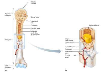
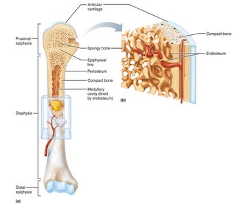
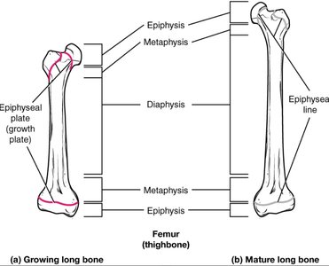
Long bones have a shaft (diaphysis), ends (epiphyses), and membranes. The diaphysis is a tubular shaft of compact bone surrounding the medullary cavity, which contains yellow bone marrow in adults. Epiphyses are the ends, consisting of compact bone externally and spongy bone internally, covered by articular cartilage. The epiphyseal line marks the site of bone growth.
Periosteum: Double-layered membrane covering external surfaces except joints; contains nerve fibers, blood vessels, and bone cells.
Endosteum: Delicate membrane covering internal bone surfaces and trabeculae of spongy bone.
Cells of Bone Tissue
Five Major Types of Bone Cells
Bone tissue contains five major cell types, each with specialized functions in bone formation, maintenance, and resorption.
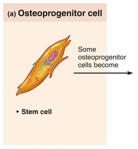
Osteoprogenitor (osteogenic) cells: Stem cells found in periosteum and endosteum; differentiate into osteoblasts or bone-lining cells.
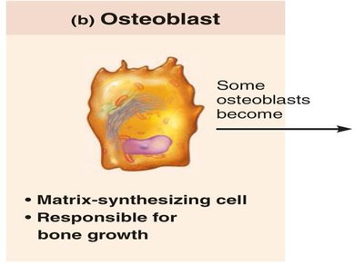
Osteoblasts: Bone-forming cells that secrete osteoid (unmineralized bone matrix); actively mitotic.
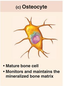
Osteocytes: Mature bone cells that maintain bone matrix and act as stress sensors.
Bone-lining cells: Flat cells on bone surfaces; help maintain matrix.
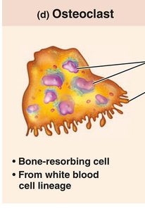
Osteoclasts: Giant, multinucleate cells responsible for bone resorption; derived from hematopoietic stem cells.
Compact vs. Spongy Bone
Structure and Location
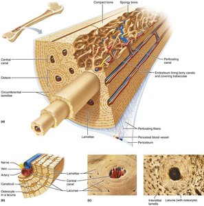
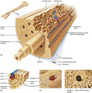
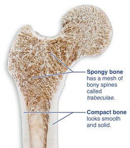
Compact bone forms the dense outer layer of bones, while spongy bone is found internally, organized along lines of stress to provide strength.
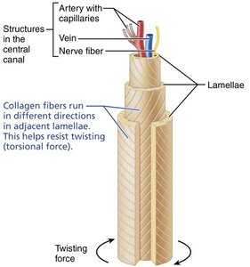
Compact Bone: Composed of osteons (Haversian systems), concentric lamellae, and canaliculi. Osteons contain a central canal for blood vessels and nerves.
Spongy Bone: Contains trabeculae, irregularly arranged lamellae, and osteocytes interconnected by canaliculi. Capillaries in endosteum supply nutrients.
Bone Markings
Categories and Functions
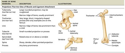
Bone markings are sites for muscle, ligament, and tendon attachment, joint formation, or passageways for blood vessels and nerves. They are classified as projections, surfaces, or depressions/openings.
Name of Bone Marking | Description | Illustrations |
|---|---|---|
Tuberosity | Large rounded projection; may be roughened | Femur, iliac spine |
Trochanter | Very large, irregularly shaped process | Femur |
Line | Narrow ridge of bone | Femur |
Tubercle | Small rounded projection or process | Humerus |
Epicondyle | Raised area above a condyle | Femur |
Spine | Sharp, slender, often pointed projection | Vertebra |
Process | Any bony prominence | Spinous process |
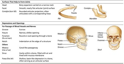
Surfaces | Description | Illustrations |
|---|---|---|
Head | Bony expansion carried on a narrow neck | Rib |
Facet | Smooth, nearly flat articular surface | Vertebra |
Condyle | Rounded articular projection | Mandible |
Depressions and Openings | Description | Illustrations |
|---|---|---|
Groove | Narrow, slitlike opening | Skull |
Fissure | Narrow, slitlike opening | Skull |
Foramen | Round or oval opening through a bone | Skull |
Notch | Indentation at the edge of a structure | Skull |
Meatus | Canal-like passageway | Skull |
Sinus | Cavity within a bone, filled with air and lined with mucous membrane | Skull |
Fossa | Shallow, basinlike depression in a bone | Skull |
Bone Development and Growth
Organic and Inorganic Components
Bones are composed of organic (cells and osteoid) and inorganic (hydroxyapatite) components. Organic components provide flexibility and strength, while inorganic components confer hardness and resistance to compression.
Organic: Osteogenic cells, osteoblasts, osteocytes, bone-lining cells, osteoclasts, and osteoid (collagen and ground substance).
Inorganic: Hydroxyapatites (calcium phosphate crystals); 65% of bone mass.
Ossification (Osteogenesis)
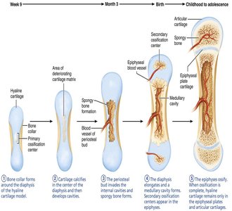
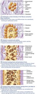
Ossification is the process of bone tissue formation, beginning in the second month of development and continuing throughout life. Bone growth occurs via two mechanisms: endochondral and intramembranous ossification.
Cartilage Growth
Appositional Growth: New matrix is laid down on the surface of cartilage by cells in the perichondrium.
Interstitial Growth: Chondrocytes within lacunae divide and secrete new matrix, expanding cartilage from within.
Growth in Length and Width of Bones
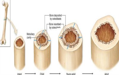
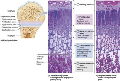
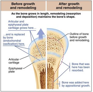
Long bones grow in length by interstitial growth at the epiphyseal plate and in width by appositional growth. The epiphyseal plate consists of five zones: resting, proliferation, hypertrophic, calcification, and ossification.
Hormonal Regulation of Bone Growth
Growth Hormone: Stimulates epiphyseal plate activity.
Thyroid Hormone: Modulates growth hormone activity.
Sex Hormones: Promote growth spurts and induce epiphyseal plate closure.
Bone Remodeling and Repair
Bone Remodeling
Bone remodeling is a continuous process involving bone deposit and resorption, coordinated by osteoblasts and osteoclasts. It is controlled by hormonal feedback and mechanical stress.
Hormonal Controls: Parathyroid hormone (PTH) and calcitonin regulate blood calcium levels.
Mechanical Stress: Bones remodel in response to stress (Wolff's law).
Fracture Classification and Repair
Fractures are classified by the position of bone ends, completeness of break, and whether the skin is penetrated. Repair involves four stages: hematoma formation, fibrocartilaginous callus formation, bony callus formation, and bone remodeling.
Stage | Description |
|---|---|
Hematoma Formation | Clotted blood forms at the fracture site. |
Fibrocartilaginous Callus Formation | Capillaries grow into hematoma; fibroblasts secrete collagen. |
Bony Callus Formation | New trabeculae form in callus; callus converts to bony callus. |
Bone Remodeling | Compact bone is laid down; structure resembles original bone. |
Additional info: Hormonal regulation and mechanical stress are essential for maintaining bone health and calcium homeostasis. Vitamin D is required for intestinal absorption of calcium.
