Indietro
Skin and Body Membranes: Structure, Function, and Clinical Aspects
Guida di studio - Note intelligenti
 Appunti personalizzati basati sui tuoi materiali, ampliati con definizioni chiave, esempi e contesto.
Body Membranes
Epithelial Membranes
Body membranes are essential structures that cover surfaces, line body cavities, and form protective sheets around organs. Epithelial membranes include the cutaneous, mucous, and serous membranes. Each type has a unique structure and function:
Cutaneous Membrane (Skin): The outer covering of the body, composed of a superficial epidermis (stratified squamous epithelium) and an underlying dermis (dense irregular connective tissue). It is exposed to air and is a dry membrane.
Mucous Membranes (Mucosae): Line all body cavities that open to the exterior (e.g., respiratory, digestive, urinary, and reproductive tracts). They consist of an epithelial layer (type varies by location) resting on a loose connective tissue membrane called the lamina propria. These membranes are moist and often adapted for absorption or secretion.
Serous Membranes (Serosae): Line closed ventral body cavities. Composed of simple squamous epithelium on a thin layer of areolar connective tissue. They occur in pairs: the parietal layer lines the cavity wall, and the visceral layer covers the organs. Serous fluid between layers reduces friction.
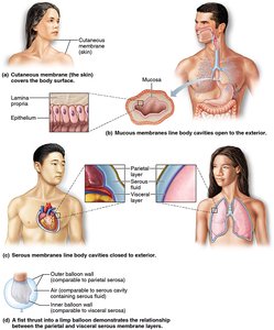
Additional info: The serous membranes are named by location: peritoneum (abdomen), pleurae (lungs), and pericardia (heart).
Connective Tissue Membranes
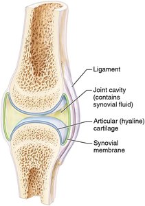
Connective tissue membranes are represented by synovial membranes, which line the fibrous capsules surrounding joints. They provide a smooth surface and secrete lubricating synovial fluid, also lining bursae and tendon sheaths to reduce friction during movement.
The Integumentary System (Skin)
Functions of the Integumentary System
The integumentary system, consisting of the skin and its appendages (sweat and oil glands, hair, and nails), performs several vital functions:
Protection: Shields deeper tissues from mechanical, chemical, microbial, UV, thermal, and desiccation damage.
Temperature Regulation: Sweat glands and blood vessels help regulate heat loss and retention.
Excretion: Sweat glands excrete urea, uric acid, and salts.
Vitamin D Synthesis: Modified cholesterol in the skin is converted to vitamin D by sunlight.
Sensory Reception: Contains receptors for touch, pressure, pain, and temperature.
Additional info: The skin's acid mantle and immune cells provide further defense against pathogens.
Structure of the Skin
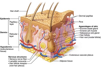
The skin is composed of two main layers: the epidermis and the dermis. Deep to the dermis is the subcutaneous tissue (hypodermis), which anchors the skin and provides insulation and nutrient storage.
Epidermis
The epidermis is a stratified squamous epithelium, avascular, and consists mainly of keratinocytes. It is organized into up to five layers (strata):
Stratum basale: Deepest layer; contains stem cells that divide to form new keratinocytes and melanocytes (produce melanin).
Stratum spinosum: Cells contain bundles of pre-keratin filaments.
Stratum granulosum: Cells flatten and accumulate keratin granules.
Stratum lucidum: Present only in thick, hairless skin (palms, soles); cells die and become clear.
Stratum corneum: Outermost, 20–30 layers of dead, keratin-filled cells; provides a tough, protective barrier.
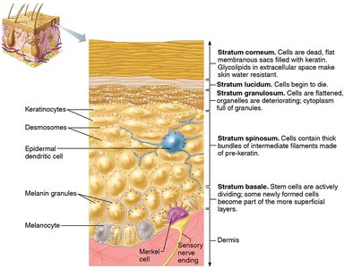
Additional info: The epidermis is renewed every 25–45 days. Melanin protects DNA from UV damage.
Dermis
The dermis is a strong, flexible connective tissue layer with two regions:
Papillary layer: Superficial; contains dermal papillae with capillaries, pain, and touch receptors. Forms fingerprints.
Reticular layer: Deep; contains dense irregular connective tissue, blood vessels, sweat and oil glands, and pressure receptors.
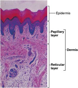
Additional info: Collagen provides toughness; elastic fibers give skin elasticity. Blood vessels help regulate temperature.
Homeostatic Imbalances: Decubitus Ulcers
Restriction of blood supply to the skin can cause cell death and ulcers, especially in bedridden patients. Decubitus ulcers (bedsores) result from prolonged pressure, leading to tissue degeneration and ulceration.
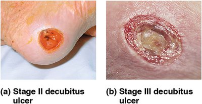
Skin Color
Skin color is determined by three pigments:
Melanin: Produced by melanocytes; provides brown, black, or yellow tones and protects against UV radiation.
Carotene: Yellow-orange pigment from diet; accumulates in the stratum corneum and hypodermis.
Hemoglobin: Oxygenated pigment in red blood cells; gives a rosy glow in light-skinned individuals.
Additional info: Cyanosis (bluish skin) indicates poor oxygenation; jaundice signals liver dysfunction; bruises result from blood leakage (hematomas).
Appendages of the Skin
Skin appendages include cutaneous glands, hair, and nails. All are derived from the epidermis but reside in the dermis.
Cutaneous Glands
Sebaceous (Oil) Glands: Secrete sebum into hair follicles; lubricates skin and hair, inhibits bacteria. Overactivity can cause acne or seborrhea.
Sweat (Sudoriferous) Glands: Two types:
Eccrine glands: Widely distributed; produce watery sweat for thermoregulation.
Apocrine glands: Found in axillary/genital areas; secrete a protein- and fat-rich fluid, which bacteria can metabolize to produce odor.
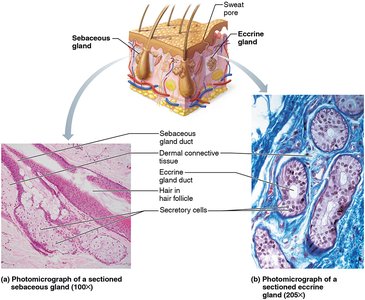
Hair and Hair Follicles
Hair is a flexible strand of dead, keratinized cells. The hair follicle is a compound structure with an inner epithelial root sheath and an outer dermal sheath. The arrector pili muscle causes hair to stand up (goosebumps). Hair color is determined by melanocyte activity.
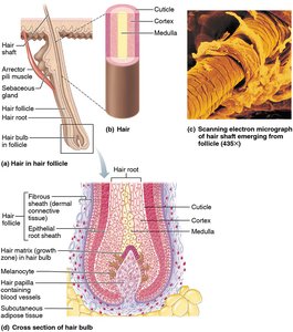
Additional info: Hair type (straight, wavy, curly) depends on the shape of the hair shaft.
Nails
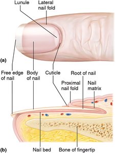
Nails are modifications of the epidermis, composed of hard keratin. The nail matrix is responsible for growth. The lunule is the white crescent at the nail base.
Homeostatic Imbalances of Skin
Infections and Allergies
Athlete’s foot: Fungal infection between toes.
Boils and carbuncles: Bacterial infection of hair follicles.
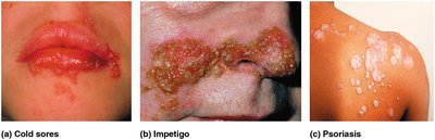
Cold sores: Viral infection (herpes simplex) causing blisters.
Contact dermatitis: Allergic reaction causing redness and blistering.
Impetigo: Bacterial infection causing crusty lesions, common in children.
Psoriasis: Autoimmune disorder with red, scaly patches.
Burns
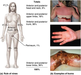
Burns are classified by depth and extent. The rule of nines estimates the percentage of body surface affected. Severity ranges from first-degree (superficial) to fourth-degree (deep tissue involvement). Major risks include fluid loss, infection, and shock.
Developmental Aspects of Skin and Body Membranes
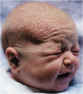
Fetal skin is covered by lanugo (fine hair) and vernix caseosa (protective, cheesy coating). At birth, skin is thin and matures with age. Adolescence brings increased oil production and possible acne. Aging leads to thinner, drier, less elastic skin, increased bruising, and hair loss or graying. Good nutrition and sun protection help delay aging effects.