 Indietro
Skin and Body Membranes: Structure, Types, and Functions
Guida di studio - Note intelligenti
 Appunti personalizzati basati sui tuoi materiali, ampliati con definizioni chiave, esempi e contesto.
Skin and Body Membranes
Introduction
The skin and body membranes are essential components of the human body, providing protection, compartmentalization, and specialized functions. This chapter explores the types, structures, and roles of body membranes, with a focus on the integumentary system.
Types and Functions of Body Membranes
Overview of Body Membranes
Cover surfaces of the body
Line body cavities
Form protective sheets around organs
There are two main types of body membranes:
Epithelial membranes
Connective tissue membranes
Epithelial Membranes
Cutaneous Membrane (Skin)
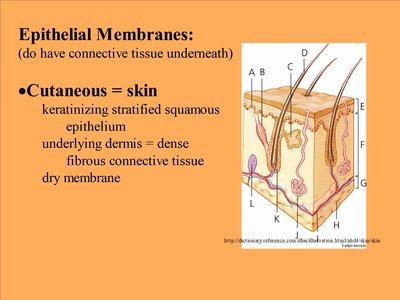
The cutaneous membrane, commonly known as the skin, is a dry membrane that covers the body surface. It consists of a keratinizing stratified squamous epithelium overlying a dense fibrous connective tissue (the dermis).
Keratinizing: The process by which cells produce keratin, a tough, protective protein.
Stratified squamous epithelium: Multiple layers of flat cells, providing durability and protection.
Dermis: Dense connective tissue beneath the epithelium.
Mucous Membranes (Mucosa)
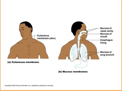
Mucous membranes line all body cavities that open to the exterior, such as the digestive, respiratory, urinary, and reproductive tracts. They consist of an epithelial layer on a loose connective tissue base called the lamina propria.
Cell makeup varies:
Stratified squamous epithelium: Found in the mouth and esophagus
Simple columnar epithelium: Found in the digestive tract
Wet membranes: Adapted for absorption and secretion
Serous Membranes (Serosa)
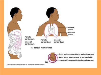
Serous membranes line body cavities that are closed to the exterior. They are composed of simple squamous epithelium on a thin layer of areolar connective tissue and occur in pairs:
Parietal layer: Lines the wall of the ventral cavity
Visceral layer: Covers the outside of organs
Serous fluid: Secreted between layers to reduce friction
Serous Membrane Names by Location
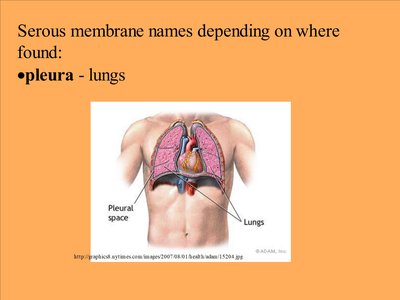
Pleura: Lungs
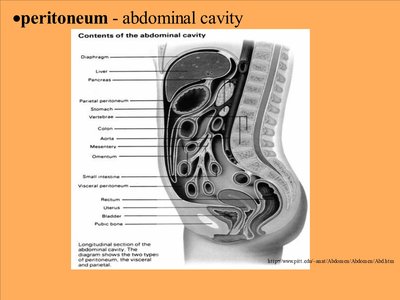
Peritoneum: Abdominal cavity
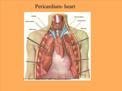
Pericardium: Heart
Connective Tissue Membranes
Synovial Membranes
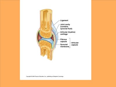
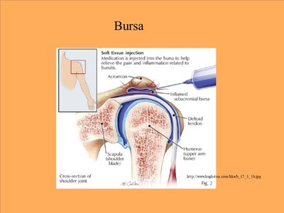
Synovial membranes are composed of soft areolar connective tissue and contain no epithelial cells. They line the fibrous capsules surrounding joints and secrete a lubricating synovial fluid. These membranes also line bursae (small sacs) and tendon sheaths, providing cushioning during muscle activity.
Secrete lubricating fluid to reduce friction in joints
Line bursae and tendon sheaths for cushioning
Functions of the Integumentary System
Protection
The skin provides a protective barrier against various types of damage:
Mechanical damage
Chemical damage
Bacterial damage
Ultraviolet (UV) radiation
Thermal damage
Desiccation (drying out)
Heat Maintenance
To lose heat: Activates sweat glands
To retain heat: Capillaries constrict
Goosebumps: Arrector pili muscles contract, making hair stand up to trap heat
Subcutaneous layer (fat): Provides insulation
Excretion and Synthesis
Excretion: Sweat glands remove urea and uric acid (nitrogenous wastes)
Synthesis of Vitamin D: Modified cholesterol molecules in the skin are converted to vitamin D by sunlight
Structure of the Skin
Major Layers
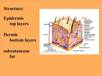
Epidermis: Top layers, composed of stratified squamous epithelium
Dermis: Bottom layers, composed of dense connective tissue
Subcutaneous (hypodermis): Fat layer for insulation
Epidermis
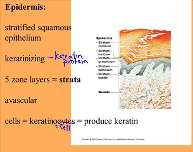
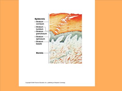
The epidermis is the outermost layer of the skin, made up of keratinizing stratified squamous epithelium. It is avascular (contains no blood vessels) and consists of five distinct layers (strata). The main cell type is the keratinocyte, which produces keratin.
Keratinocytes: Cells that produce keratin protein
Strata: Five layers from deep to superficial
Stratum Basale (Deepest Layer)
Also called stratum germinativum
Actively mitotic: Cells divide rapidly, pushing daughter cells upwards
Closest to the dermis and receives nourishment from it
Contains melanocytes that produce melanin (pigment responsible for skin color)
Sun exposure increases melanin production, which protects DNA from UV damage
Freckles and moles are concentrated areas of melanin
Additional info: The skin is the largest organ of the body and plays a critical role in homeostasis, sensory perception, and immune defense.
